2C9E

Peridinin-chlorophyll a protein, high-salt form


Experimental Data Snapshot

  • Method: X-RAY DIFFRACTION
  • Resolution: 2.10 Å
  • R-Value Free: 0.219 
  • R-Value Work: 0.174 
  • R-Value Observed: 0.174 

wwPDB Validation   3D Report Full Report


Ligand Structure Quality Assessment 


This is version 2.0 of the entry. See complete history


Literature

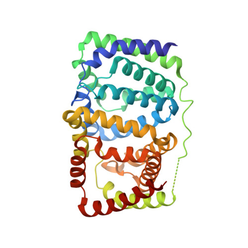
X-Ray Structure of the High-Salt Form of the Peridinin-Chlorophyll A-Protein from the Dinoflagellate Amphidinium Carterae: Modulation of the Spectral Properties of Pigments by the Protein Environment.

Schulte, T.Sharples, F.P.Hiller, R.G.Hofmann, E.

(2009) Biochemistry 48: 4466

  • DOI: https://doi.org/10.1021/bi802320q
  • Primary Citation of Related Structures:  
    2C9E

  • PubMed Abstract: 

    Light-harvesting complexes have evolved into very different structures but fulfill the same function, efficient harvesting of solar energy. In these complexes, pigments are fine-tuned and properly arranged to gather incoming photons. In the photosynthetic dinoflagellate Amphidinium carterae, two variants of the soluble light-harvesting complex PCP have been found [main form PCP (MFPCP) and high-salt PCP (HSPCP)], which show small variations in their pigment arrangement and tuning mechanisms. This feature makes them ideal models for studying pigment-protein interactions. Here we present the X-ray structure of the monomeric HSPCP determined at 2.1 A resolution and compare it to the structure of trimeric MFPCP. Despite the high degree of structural similarity (rmsd C(alpha)-C(alpha) of 1.89 A), the sequence variations lead to a changed overall pigment composition which includes the loss of two carotenoid molecules and a dramatic rearrangement of the chlorophyll phytol chains and of internal lipid molecules. On the basis of a detailed structural comparison, we favor a macrocycle geometry distortion of the chlorophylls rather than an electrostatic effect to explain energetic splitting of the chlorophyll a Q(y) bands [Ilagan, R. P. (2006) Biochemistry 45, 14052-14063]. Our analysis supports their assignment of peridinin 611* as the single blue-shifted peridinin in HSPCP but also highlights another electrostatic feature due to glutamate 202 which could add to the observed binding site asymmetry of the 611*/621* peridinin pair.


  • Organizational Affiliation

    Biophysics, Department of Biology and Biotechnology, Ruhr-University Bochum, D-44780 Bochum, Germany.


Macromolecules
Find similar proteins by:  (by identity cutoff)  |  3D Structure
Entity ID: 1
MoleculeChains Sequence LengthOrganismDetailsImage
PERIDININ-CHLOROPHYLL A PROTEIN327Amphidinium carteraeMutation(s): 0 
UniProt
Find proteins for O76183 (Amphidinium carterae)
Explore O76183 
Go to UniProtKB:  O76183
Entity Groups  
Sequence Clusters30% Identity50% Identity70% Identity90% Identity95% Identity100% Identity
UniProt GroupO76183
Sequence Annotations
Expand
  • Reference Sequence
Small Molecules
Ligands 4 Unique
IDChains Name / Formula / InChI Key2D Diagram3D Interactions
DGD
Query on DGD

Download Ideal Coordinates CCD File 
G [auth A],
K [auth A]
DIGALACTOSYL DIACYL GLYCEROL (DGDG)
C51 H96 O15
LDQFLSUQYHBXSX-HXXRYREZSA-N
CLA
Query on CLA

Download Ideal Coordinates CCD File 
B [auth A],
C [auth A]
CHLOROPHYLL A
C55 H72 Mg N4 O5
ATNHDLDRLWWWCB-AENOIHSZSA-M
PID
Query on PID

Download Ideal Coordinates CCD File 
D [auth A]
E [auth A]
F [auth A]
H [auth A]
I [auth A]
D [auth A],
E [auth A],
F [auth A],
H [auth A],
I [auth A],
J [auth A]
PERIDININ
C39 H50 O7
UYRDHEJRPVSJFM-FROCQLDGSA-N
MG
Query on MG

Download Ideal Coordinates CCD File 
L [auth A]MAGNESIUM ION
Mg
JLVVSXFLKOJNIY-UHFFFAOYSA-N
Experimental Data & Validation

Experimental Data

  • Method: X-RAY DIFFRACTION
  • Resolution: 2.10 Å
  • R-Value Free: 0.219 
  • R-Value Work: 0.174 
  • R-Value Observed: 0.174 
  • Space Group: P 1 21 1
Unit Cell:
Length ( Å )Angle ( ˚ )
a = 46.576α = 90
b = 62.673β = 100.89
c = 63.345γ = 90
Software Package:
Software NamePurpose
CNSrefinement
XDSdata reduction
XSCALEdata scaling
MOLREPphasing

Structure Validation

View Full Validation Report


Ligand Structure Quality Assessment 


Entry History 

Deposition Data

Revision History  (Full details and data files)

  • Version 1.0: 2007-02-20
    Type: Initial release
  • Version 1.1: 2012-08-08
    Changes: Atomic model, Derived calculations, Non-polymer description, Version format compliance
  • Version 2.0: 2023-12-13
    Changes: Data collection, Database references, Derived calculations, Non-polymer description, Other, Refinement description