2AJ4

Crystal structure of Saccharomyces cerevisiae Galactokinase in complex with galactose and Mg:AMPPNP


Experimental Data Snapshot

  • Method: X-RAY DIFFRACTION
  • Resolution: 2.40 Å
  • R-Value Free: 0.205 
  • R-Value Work: 0.172 
  • R-Value Observed: 0.174 

wwPDB Validation   3D Report Full Report


Ligand Structure Quality Assessment 


This is version 1.4 of the entry. See complete history


Literature

Molecular structure of Saccharomyces cerevisiae Gal1p, a bifunctional galactokinase and transcriptional inducer

Thoden, J.B.Sellick, C.A.Timson, D.J.Reece, R.J.Holden, H.M.

(2005) J Biol Chem 280: 36905-36911

  • DOI: https://doi.org/10.1074/jbc.M508446200
  • Primary Citation of Related Structures:  
    2AJ4

  • PubMed Abstract: 

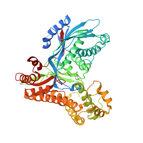
    Gal1p of Saccharomyces cerevisiae is capable of performing two independent cellular functions. First, it is a key enzyme in the Leloir pathway for galactose metabolism where it catalyzes the conversion of alpha-d-galactose to galactose 1-phosphate. Second, it has the capacity to induce the transcription of the yeast GAL genes in response to the organism being challenged with galactose as the sole source of carbon. This latter function is normally performed by a highly related protein, Gal3p, but in its absence Gal1p can induce transcription, albeit inefficiently, both in vivo and in vitro. Here we report the x-ray structure of Gal1p in complex with alpha-d-galactose and Mg-adenosine 5'-(beta,gamma-imido)triphosphate (AMPPNP) determined to 2.4 Angstrom resolution. Overall, the enzyme displays a marked bilobal appearance with the active site being wedged between distinct N- and C-terminal domains. Despite being considerably larger than other galactokinases, Gal1p shares a similar molecular architecture with these enzymes as well as with other members of the GHMP superfamily. The extraordinary levels of similarity between Gal1p and Gal3p ( approximately 70% amino acid identity and approximately 90% similarity) have allowed a model for Gal3p to be constructed. By identifying the locations of mutations of Gal3p that result in altered transcriptional properties, we suggest potential models for Gal3p function and mechanisms for its interaction with the transcriptional inhibitor Gal80p. The GAL genetic switch has long been regarded as a paradigm for the control of gene expression in eukaryotes. Understanding the manner in which two of the proteins that function in transcriptional regulation interact with one another is an important step in determining the overall molecular mechanism of this switch.


  • Organizational Affiliation

    Department of Biochemistry, University of Wisconsin, Madison, Wisconsin 53706, USA.


Macromolecules
Find similar proteins by:  (by identity cutoff)  |  3D Structure
Entity ID: 1
MoleculeChains Sequence LengthOrganismDetailsImage
Galactokinase
A, B
548Saccharomyces cerevisiaeMutation(s): 0 
Gene Names: GAL1
EC: 2.7.1.6
UniProt
Find proteins for P04385 (Saccharomyces cerevisiae (strain ATCC 204508 / S288c))
Explore P04385 
Go to UniProtKB:  P04385
Entity Groups  
Sequence Clusters30% Identity50% Identity70% Identity90% Identity95% Identity100% Identity
UniProt GroupP04385
Sequence Annotations
Expand
  • Reference Sequence
Small Molecules
Ligands 4 Unique
IDChains Name / Formula / InChI Key2D Diagram3D Interactions
ANP
Query on ANP

Download Ideal Coordinates CCD File 
F [auth A],
J [auth B]
PHOSPHOAMINOPHOSPHONIC ACID-ADENYLATE ESTER
C10 H17 N6 O12 P3
PVKSNHVPLWYQGJ-KQYNXXCUSA-N
GLA
Query on GLA

Download Ideal Coordinates CCD File 
C [auth A],
G [auth B]
alpha-D-galactopyranose
C6 H12 O6
WQZGKKKJIJFFOK-PHYPRBDBSA-N
CL
Query on CL

Download Ideal Coordinates CCD File 
E [auth A],
I [auth B]
CHLORIDE ION
Cl
VEXZGXHMUGYJMC-UHFFFAOYSA-M
MG
Query on MG

Download Ideal Coordinates CCD File 
D [auth A],
H [auth B]
MAGNESIUM ION
Mg
JLVVSXFLKOJNIY-UHFFFAOYSA-N
Experimental Data & Validation

Experimental Data

  • Method: X-RAY DIFFRACTION
  • Resolution: 2.40 Å
  • R-Value Free: 0.205 
  • R-Value Work: 0.172 
  • R-Value Observed: 0.174 
  • Space Group: P 1 21 1
Unit Cell:
Length ( Å )Angle ( ˚ )
a = 63.3α = 90
b = 85β = 96.7
c = 112.3γ = 90
Software Package:
Software NamePurpose
PROTEUM PLUSdata collection
SAINTdata reduction
EPMRphasing
TNTrefinement
PROTEUM PLUSdata reduction
SAINTdata scaling

Structure Validation

View Full Validation Report


Ligand Structure Quality Assessment 


Entry History 

Deposition Data

Revision History  (Full details and data files)

  • Version 1.0: 2005-08-30
    Type: Initial release
  • Version 1.1: 2008-04-30
    Changes: Version format compliance
  • Version 1.2: 2011-07-13
    Changes: Version format compliance
  • Version 1.3: 2020-07-29
    Type: Remediation
    Reason: Carbohydrate remediation
    Changes: Data collection, Database references, Derived calculations, Structure summary
  • Version 1.4: 2023-08-23
    Changes: Data collection, Database references, Refinement description, Structure summary