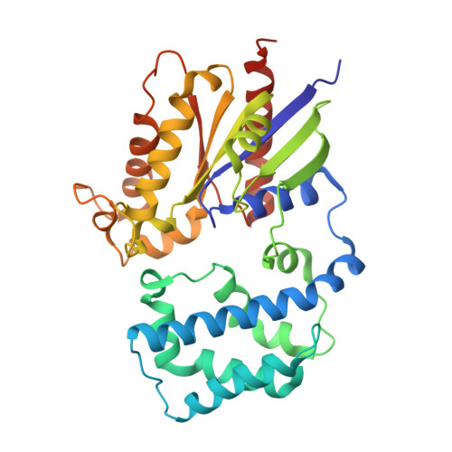
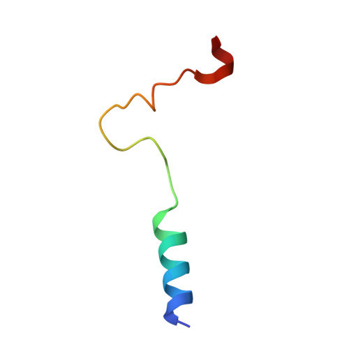
Structure-based Protocol for Identifying Mutations that Enhance Protein-Protein Binding Affinities.
Sammond, D.W., Eletr, Z.M., Purbeck, C., Kimple, R.J., Siderovski, D.P., Kuhlman, B.(2007) J Mol Biol 371: 1392-1404
- PubMed: 17603074
- DOI: https://doi.org/10.1016/j.jmb.2007.05.096
- Primary Citation of Related Structures:
2OM2 - PubMed Abstract:
The ability to manipulate protein binding affinities is important for the development of proteins as biosensors, industrial reagents, and therapeutics. We have developed a structure-based method to rationally predict single mutations at protein-protein interfaces that enhance binding affinities. The protocol is based on the premise that increasing buried hydrophobic surface area and/or reducing buried hydrophilic surface area will generally lead to enhanced affinity if large steric clashes are not introduced and buried polar groups are not left without a hydrogen bond partner. The procedure selects affinity enhancing point mutations at the protein-protein interface using three criteria: (1) the mutation must be from a polar amino acid to a non-polar amino acid or from a non-polar amino acid to a larger non-polar amino acid, (2) the free energy of binding as calculated with the Rosetta protein modeling program should be more favorable than the free energy of binding calculated for the wild-type complex and (3) the mutation should not be predicted to significantly destabilize the monomers. The performance of the computational protocol was experimentally tested on two separate protein complexes; Galpha(i1) from the heterotrimeric G-protein system bound to the RGS14 GoLoco motif, and the E2, UbcH7, bound to the E3, E6AP from the ubiquitin pathway. Twelve single-site mutations that were predicted to be stabilizing were synthesized and characterized in the laboratory. Nine of the 12 mutations successfully increased binding affinity with five of these increasing binding by over 1.0 kcal/mol. To further assess our approach we searched the literature for point mutations that pass our criteria and have experimentally determined binding affinities. Of the eight mutations identified, five were accurately predicted to increase binding affinity, further validating the method as a useful tool to increase protein-protein binding affinities.
Organizational Affiliation:
Department of Biochemistry and Biophysics, University of North Carolina at Chapel Hill, Chapel Hill, NC 27599-7260, USA.