2IDX

Structure of Human ATP:Cobalamin adenosyltransferase bound to ATP.


Experimental Data Snapshot

  • Method: X-RAY DIFFRACTION
  • Resolution: 2.50 Å
  • R-Value Free: 0.251 
  • R-Value Work: 0.197 
  • R-Value Observed: 0.200 

wwPDB Validation   3D Report Full Report


Ligand Structure Quality Assessment 


This is version 1.4 of the entry. See complete history


Literature

Structure of ATP-Bound Human ATP:Cobalamin Adenosyltransferase.

Schubert, H.L.Hill, C.P.

(2006) Biochemistry 45: 15188-15196

  • DOI: https://doi.org/10.1021/bi061396f
  • Primary Citation of Related Structures:  
    2IDX

  • PubMed Abstract: 

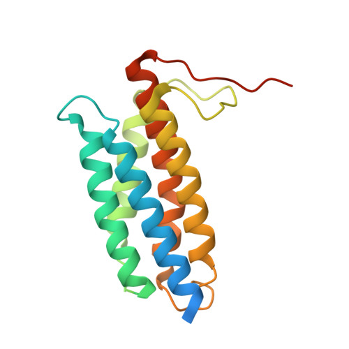
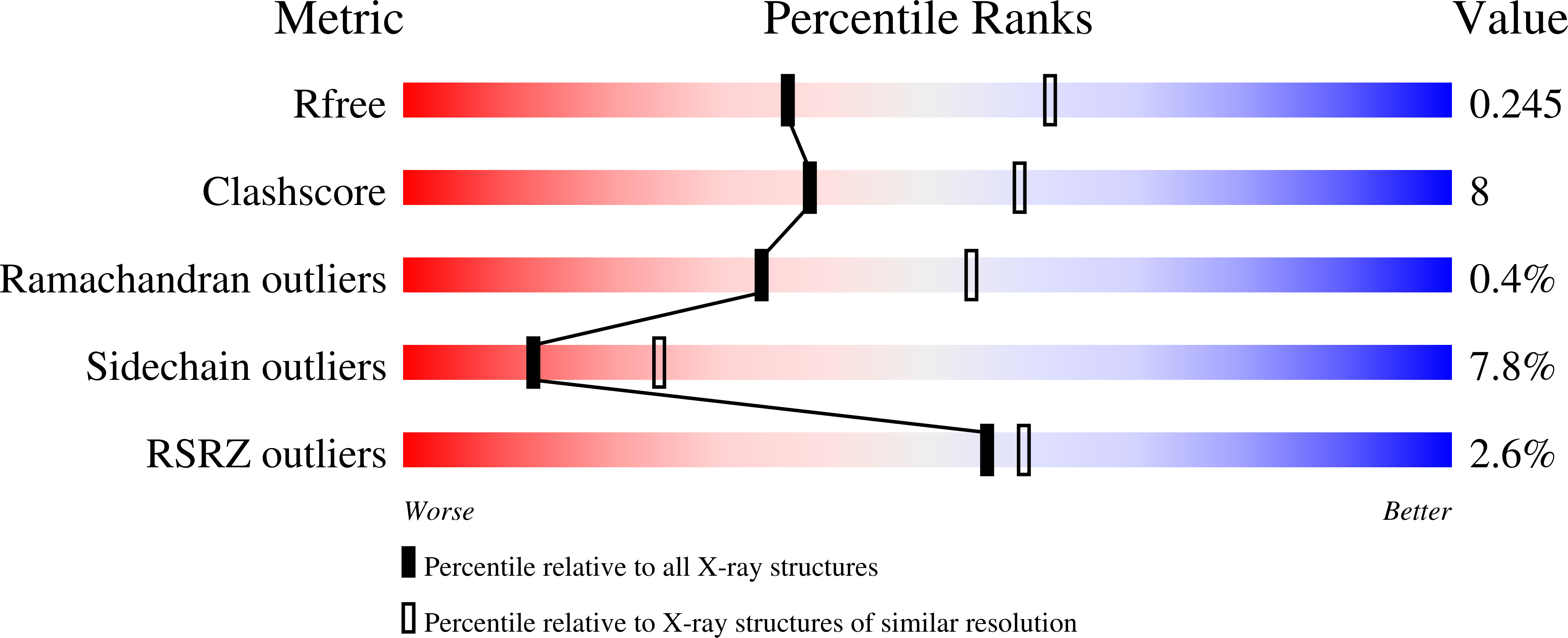
    Mutations in the gene encoding human ATP:cobalamin adenosyltransferase (hATR) can result in the metabolic disorder known as methylmalonic aciduria (MMA). This enzyme catalyzes the final step in the conversion of cyanocobalamin (vitamin B12) to the essential human cofactor adenosylcobalamin. Here we present the 2.5 A crystal structure of ATP bound to hATR refined to an Rfree value of 25.2%. The enzyme forms a tightly associated trimer, where the monomer comprises a five-helix bundle and the active sites lie on the subunit interfaces. Only two of the three active sites within the trimer contain the bound ATP substrate, thereby providing examples of apo- and substrate-bound-active sites within the same crystal structure. Comparison of the empty and occupied sites indicates that twenty residues at the enzyme's N-terminus become ordered upon binding of ATP to form a novel ATP-binding site and an extended cleft that likely binds cobalamin. The structure explains the role of 20 invariant residues; six are involved in ATP binding, including Arg190, which hydrogen bonds to ATP atoms on both sides of the scissile bond. Ten of the hydrogen bonds are required for structural stability, and four are in positions to interact with cobalamin. The structure also reveals how the point mutations that cause MMA are deficient in these functions.


  • Organizational Affiliation

    Department of Biochemistry, University of Utah, Salt Lake City, Utah 84112-5650, USA. heidi@biochem.utah.edu


Macromolecules
Find similar proteins by:  (by identity cutoff)  |  3D Structure
Entity ID: 1
MoleculeChains Sequence LengthOrganismDetailsImage
Cob(I)yrinic acid a,c-diamide adenosyltransferase
A, B, C
196Homo sapiensMutation(s): 0 
Gene Names: MMAB
EC: 2.5.1.17
UniProt & NIH Common Fund Data Resources
Find proteins for Q96EY8 (Homo sapiens)
Explore Q96EY8 
Go to UniProtKB:  Q96EY8
PHAROS:  Q96EY8
GTEx:  ENSG00000139428 
Entity Groups  
Sequence Clusters30% Identity50% Identity70% Identity90% Identity95% Identity100% Identity
UniProt GroupQ96EY8
Sequence Annotations
Expand
  • Reference Sequence
Small Molecules
Ligands 4 Unique
IDChains Name / Formula / InChI Key2D Diagram3D Interactions
ATP
Query on ATP

Download Ideal Coordinates CCD File 
H [auth A],
K [auth B]
ADENOSINE-5'-TRIPHOSPHATE
C10 H16 N5 O13 P3
ZKHQWZAMYRWXGA-KQYNXXCUSA-N
SO4
Query on SO4

Download Ideal Coordinates CCD File 
D [auth A],
I [auth B],
L [auth C]
SULFATE ION
O4 S
QAOWNCQODCNURD-UHFFFAOYSA-L
CL
Query on CL

Download Ideal Coordinates CCD File 
E [auth A]CHLORIDE ION
Cl
VEXZGXHMUGYJMC-UHFFFAOYSA-M
MG
Query on MG

Download Ideal Coordinates CCD File 
F [auth A],
G [auth A],
J [auth B],
M [auth C]
MAGNESIUM ION
Mg
JLVVSXFLKOJNIY-UHFFFAOYSA-N
Experimental Data & Validation

Experimental Data

  • Method: X-RAY DIFFRACTION
  • Resolution: 2.50 Å
  • R-Value Free: 0.251 
  • R-Value Work: 0.197 
  • R-Value Observed: 0.200 
  • Space Group: P 31 2 1
Unit Cell:
Length ( Å )Angle ( ˚ )
a = 111.238α = 90
b = 111.238β = 90
c = 115.526γ = 120
Software Package:
Software NamePurpose
DENZOdata reduction
SCALEPACKdata scaling
PHASERphasing
REFMACrefinement
PDB_EXTRACTdata extraction

Structure Validation

View Full Validation Report


Ligand Structure Quality Assessment 


Entry History 

Deposition Data

Revision History  (Full details and data files)

  • Version 1.0: 2007-02-06
    Type: Initial release
  • Version 1.1: 2008-05-01
    Changes: Version format compliance
  • Version 1.2: 2011-07-13
    Changes: Version format compliance
  • Version 1.3: 2017-10-18
    Changes: Refinement description
  • Version 1.4: 2023-08-30
    Changes: Data collection, Database references, Derived calculations, Refinement description