2C4H

Torpedo californica acetylcholinesterase in complex with 500mM acetylthiocholine


Experimental Data Snapshot

  • Method: X-RAY DIFFRACTION
  • Resolution: 2.15 Å
  • R-Value Free: 0.209 
  • R-Value Work: 0.169 
  • R-Value Observed: 0.169 

wwPDB Validation   3D Report Full Report


Ligand Structure Quality Assessment 


This is version 2.3 of the entry. See complete history


Literature

Structural insights into substrate traffic and inhibition in acetylcholinesterase.

Colletier, J.P.Fournier, D.Greenblatt, H.M.Stojan, J.Sussman, J.L.Zaccai, G.Silman, I.Weik, M.

(2006) EMBO J 25: 2746-2756

  • DOI: https://doi.org/10.1038/sj.emboj.7601175
  • Primary Citation of Related Structures:  
    2C4H, 2C58, 2C5F, 2C5G

  • PubMed Abstract: 

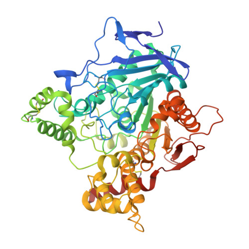
    Acetylcholinesterase (AChE) terminates nerve-impulse transmission at cholinergic synapses by rapid hydrolysis of the neurotransmitter, acetylcholine. Substrate traffic in AChE involves at least two binding sites, the catalytic and peripheral anionic sites, which have been suggested to be allosterically related and involved in substrate inhibition. Here, we present the crystal structures of Torpedo californica AChE complexed with the substrate acetylthiocholine, the product thiocholine and a nonhydrolysable substrate analogue. These structures provide a series of static snapshots of the substrate en route to the active site and identify, for the first time, binding of substrate and product at both the peripheral and active sites. Furthermore, they provide structural insight into substrate inhibition in AChE at two different substrate concentrations. Our structural data indicate that substrate inhibition at moderate substrate concentration is due to choline exit being hindered by a substrate molecule bound at the peripheral site. At the higher concentration, substrate inhibition arises from prevention of exit of acetate due to binding of two substrate molecules within the active-site gorge.


  • Organizational Affiliation

    Laboratoire de Biophysique Moléculaire, Institut de Biologie Structurale (CEA/CNRS/UJF), Grenoble Cedex, France.


Macromolecules
Find similar proteins by:  (by identity cutoff)  |  3D Structure
Entity ID: 1
MoleculeChains Sequence LengthOrganismDetailsImage
ACETYLCHOLINESTERASE537Tetronarce californicaMutation(s): 0 
EC: 3.1.1.7
UniProt
Find proteins for P04058 (Tetronarce californica)
Explore P04058 
Go to UniProtKB:  P04058
Entity Groups  
Sequence Clusters30% Identity50% Identity70% Identity90% Identity95% Identity100% Identity
UniProt GroupP04058
Sequence Annotations
Expand
  • Reference Sequence
Small Molecules
Ligands 6 Unique
IDChains Name / Formula / InChI Key2D Diagram3D Interactions
NAG
Query on NAG

Download Ideal Coordinates CCD File 
B [auth A],
C [auth A]
2-acetamido-2-deoxy-beta-D-glucopyranose
C8 H15 N O6
OVRNDRQMDRJTHS-FMDGEEDCSA-N
AT3
Query on AT3

Download Ideal Coordinates CCD File 
F [auth A],
G [auth A]
ACETYLTHIOCHOLINE
C7 H16 N O S
GFFIJCYHQYHUHB-UHFFFAOYSA-N
PGE
Query on PGE

Download Ideal Coordinates CCD File 
D [auth A],
E [auth A]
TRIETHYLENE GLYCOL
C6 H14 O4
ZIBGPFATKBEMQZ-UHFFFAOYSA-N
ACT
Query on ACT

Download Ideal Coordinates CCD File 
T [auth A]ACETATE ION
C2 H3 O2
QTBSBXVTEAMEQO-UHFFFAOYSA-M
ACE
Query on ACE

Download Ideal Coordinates CCD File 
H [auth A]ACETYL GROUP
C2 H4 O
IKHGUXGNUITLKF-UHFFFAOYSA-N
CL
Query on CL

Download Ideal Coordinates CCD File 
I [auth A]
J [auth A]
K [auth A]
L [auth A]
M [auth A]
I [auth A],
J [auth A],
K [auth A],
L [auth A],
M [auth A],
N [auth A],
O [auth A],
P [auth A],
Q [auth A],
R [auth A],
S [auth A]
CHLORIDE ION
Cl
VEXZGXHMUGYJMC-UHFFFAOYSA-M
Experimental Data & Validation

Experimental Data

  • Method: X-RAY DIFFRACTION
  • Resolution: 2.15 Å
  • R-Value Free: 0.209 
  • R-Value Work: 0.169 
  • R-Value Observed: 0.169 
  • Space Group: P 31 2 1
Unit Cell:
Length ( Å )Angle ( ˚ )
a = 111.99α = 90
b = 111.99β = 90
c = 137.12γ = 120
Software Package:
Software NamePurpose
CNSrefinement
XDSdata reduction
XSCALEdata scaling
CNSphasing

Structure Validation

View Full Validation Report


Ligand Structure Quality Assessment 


Entry History 

Deposition Data

Revision History  (Full details and data files)

  • Version 1.0: 2006-06-14
    Type: Initial release
  • Version 1.1: 2011-05-07
    Changes: Version format compliance
  • Version 1.2: 2011-07-13
    Changes: Version format compliance
  • Version 2.0: 2018-07-18
    Changes: Advisory, Atomic model, Data collection, Database references, Derived calculations, Non-polymer description, Structure summary
  • Version 2.1: 2019-04-03
    Changes: Data collection, Derived calculations, Experimental preparation
  • Version 2.2: 2020-07-29
    Type: Remediation
    Reason: Carbohydrate remediation
    Changes: Data collection, Derived calculations, Other, Structure summary
  • Version 2.3: 2023-12-13
    Changes: Data collection, Database references, Refinement description, Structure summary