1YUK

The crystal structure of the PSI/Hybrid domain/ I-EGF1 segment from the human integrin beta2 at 1.8 resolution


Experimental Data Snapshot

  • Method: X-RAY DIFFRACTION
  • Resolution: 1.80 Å
  • R-Value Free: 0.253 
  • R-Value Work: 0.234 
  • R-Value Observed: 0.234 

wwPDB Validation   3D Report Full Report


Ligand Structure Quality Assessment 


This is version 2.0 of the entry. See complete history

Re-refinement Note

A newer entry is available that reflects an alternative modeling of the original data: 5E6V


Literature

The Crystal Structure of the Plexin-Semaphorin-Integrin Domain/Hybrid Domain/I-EGF1 Segment from the Human Integrin {beta}2 Subunit at 1.8-A Resolution

Shi, M.Sundramurthy, K.Liu, B.Tan, S.M.Law, S.K.Lescar, J.

(2005) J Biol Chem 280: 30586-30593

  • DOI: https://doi.org/10.1074/jbc.M502525200
  • Primary Citation of Related Structures:  
    1YUK

  • PubMed Abstract: 

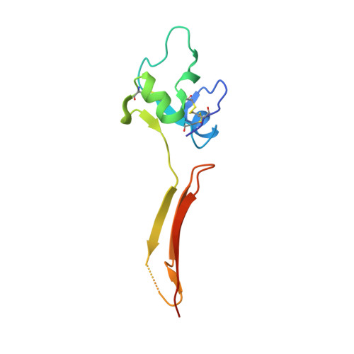
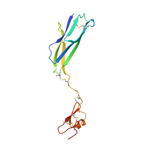
    Integrins are modular (alphabeta) heterodimeric proteins that mediate cell adhesion and convey signals across the plasma membrane. Interdomain motions play a key role in signal transduction by propagating structural changes through the molecule, thus controlling the activation state and adhesive properties of the integrin. We expressed a soluble fragment of the human integrin beta2 subunit comprising the plexin-semaphorin-integrin domain (PSI)/hybrid domain/I-EGF1 fragment and present its crystal structure at 1.8-A resolution. The structure reveals an elongated molecule with a rigid architecture stabilized by nine disulfide bridges. The PSI domain is located centrally and participates in the formation of extended interfaces with the hybrid domain and I-EGF1 domains, respectively. The hybrid domain/PSI interface involves the burial of an Arg residue, and contacts between PSI and I-EGF1 are mainly mediated by well conserved Arg and Trp residues. Conservation of key interacting residues across the various integrin beta subunits sequences suggests that our structure represents a good model for the entire integrin family. Superposition with the integrin beta3 receptor in its bent conformation suggests that an articulation point is present at the linkage between its I-EGF1 and I-EGF2 modules and underlines the importance of this region for the control of integrin-mediated cell adhesion.


  • Organizational Affiliation

    School of Biological Sciences, Nanyang Technological University, 60 Nanyang Drive, Singapore 637551, Singapore.


Macromolecules
Find similar proteins by:  (by identity cutoff)  |  3D Structure
Entity ID: 1
MoleculeChains Sequence LengthOrganismDetailsImage
Integrin beta-2 A chain103Homo sapiensMutation(s): 0 
UniProt & NIH Common Fund Data Resources
Find proteins for P05107 (Homo sapiens)
Explore P05107 
Go to UniProtKB:  P05107
PHAROS:  P05107
GTEx:  ENSG00000160255 
Entity Groups  
Sequence Clusters30% Identity50% Identity70% Identity90% Identity95% Identity100% Identity
UniProt GroupP05107
Sequence Annotations
Expand
  • Reference Sequence
Find similar proteins by:  (by identity cutoff)  |  3D Structure
Entity ID: 2
MoleculeChains Sequence LengthOrganismDetailsImage
Integrin beta-2 B chain120Homo sapiensMutation(s): 0 
UniProt & NIH Common Fund Data Resources
Find proteins for P05107 (Homo sapiens)
Explore P05107 
Go to UniProtKB:  P05107
PHAROS:  P05107
GTEx:  ENSG00000160255 
Entity Groups  
Sequence Clusters30% Identity50% Identity70% Identity90% Identity95% Identity100% Identity
UniProt GroupP05107
Sequence Annotations
Expand
  • Reference Sequence
Small Molecules
Ligands 1 Unique
IDChains Name / Formula / InChI Key2D Diagram3D Interactions
NDG
Query on NDG

Download Ideal Coordinates CCD File 
C [auth A],
D [auth B]
2-acetamido-2-deoxy-alpha-D-glucopyranose
C8 H15 N O6
OVRNDRQMDRJTHS-PVFLNQBWSA-N
Experimental Data & Validation

Experimental Data

  • Method: X-RAY DIFFRACTION
  • Resolution: 1.80 Å
  • R-Value Free: 0.253 
  • R-Value Work: 0.234 
  • R-Value Observed: 0.234 
  • Space Group: P 1 21 1
Unit Cell:
Length ( Å )Angle ( ˚ )
a = 58.6α = 90
b = 31.818β = 91.2
c = 74.95γ = 90
Software Package:
Software NamePurpose
MOSFLMdata reduction
SCALAdata scaling
SOLVEphasing
CNSrefinement
CCP4data scaling

Structure Validation

View Full Validation Report


Ligand Structure Quality Assessment 


Entry History 

Deposition Data

Revision History  (Full details and data files)

  • Version 1.0: 2005-07-19
    Type: Initial release
  • Version 1.1: 2008-04-30
    Changes: Version format compliance
  • Version 1.2: 2011-07-13
    Changes: Version format compliance
  • Version 2.0: 2020-07-29
    Type: Remediation
    Reason: Carbohydrate remediation
    Changes: Atomic model, Data collection, Database references, Derived calculations, Structure summary