1YKB

Crystal Structure of Insect Cell Expressed IL-22


Experimental Data Snapshot

  • Method: X-RAY DIFFRACTION
  • Resolution: 2.60 Å
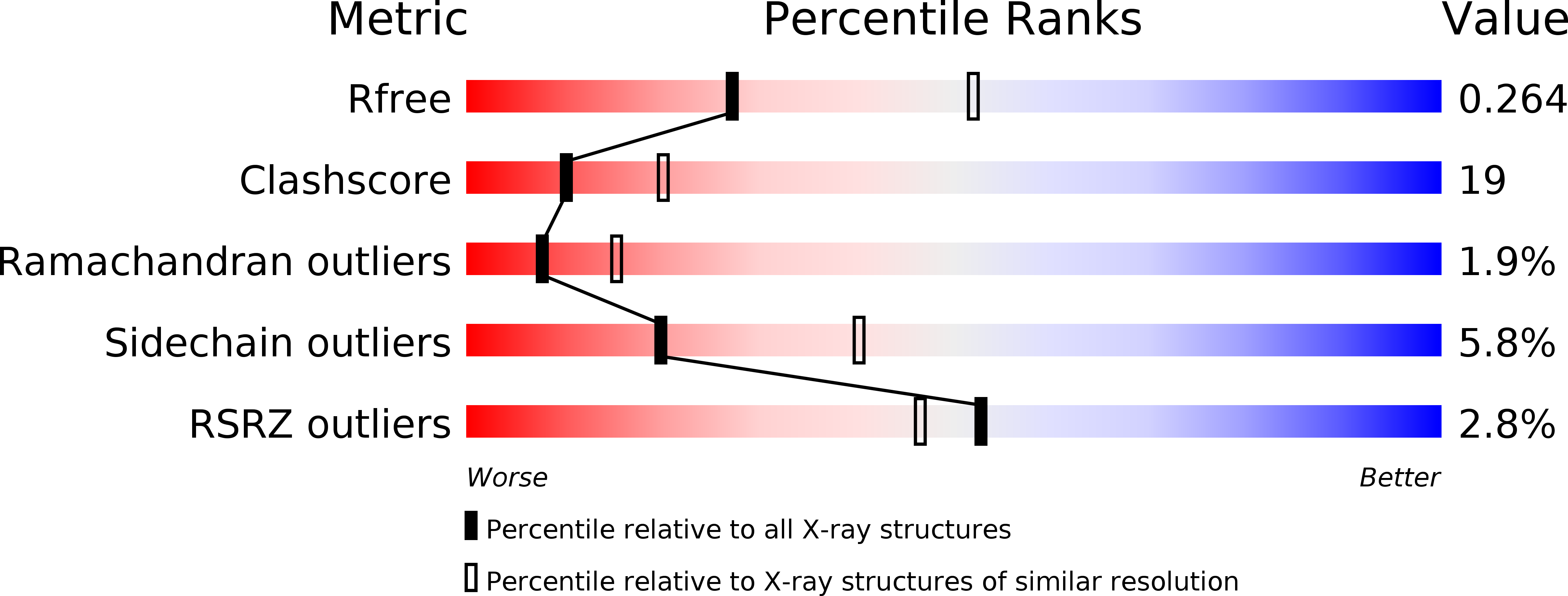
  • R-Value Free: 0.265 
  • R-Value Work: 0.232 

wwPDB Validation   3D Report Full Report


Ligand Structure Quality Assessment 


This is version 2.0 of the entry. See complete history


Literature

Structure of insect-cell-derived IL-22.

Xu, T.Logsdon, N.J.Walter, M.R.

(2005) Acta Crystallogr D Biol Crystallogr 61: 942-950

  • DOI: https://doi.org/10.1107/S0907444905009601
  • Primary Citation of Related Structures:  
    1YKB

  • PubMed Abstract: 

    The crystal structure of interleukin-22 expressed in Drosophila melanogaster S2 cells (IL-22(Dm)) has been determined at 2.6 A resolution. IL-22(Dm) crystals contain six molecules in the asymmetric unit. Comparison of IL-22(Dm) and IL-22(Ec) (interleukin-22 produced in Escherichia coli) structures reveals that N-linked glycosylation causes only minor structural changes to the cytokine. However, 1-4 A main-chain differences are observed between the six IL-22(Dm) monomers at regions corresponding to the IL-22R1 and IL-10R2 binding sites. The structure of the carbohydrate and the conformational variation of IL22(Dm) provide new insights into IL-22 receptor recognition.


  • Organizational Affiliation

    Center for Biophysical Sciences and Engineering, University of Alabama at Birmingham, Birmingham, AL 35294, USA.


Macromolecules
Find similar proteins by:  (by identity cutoff)  |  3D Structure
Entity ID: 1
MoleculeChains Sequence LengthOrganismDetailsImage
PROTEIN (Interleukin-22)
A, B, C, D, E
A, B, C, D, E, F
142Homo sapiensMutation(s): 0 
Gene Names: IL22ILTIF
UniProt & NIH Common Fund Data Resources
Find proteins for Q9GZX6 (Homo sapiens)
Explore Q9GZX6 
Go to UniProtKB:  Q9GZX6
PHAROS:  Q9GZX6
GTEx:  ENSG00000127318 
Entity Groups  
Sequence Clusters30% Identity50% Identity70% Identity90% Identity95% Identity100% Identity
UniProt GroupQ9GZX6
Sequence Annotations
Expand
  • Reference Sequence
Oligosaccharides

Help

Entity ID: 2
MoleculeChains Length2D Diagram Glycosylation3D Interactions
2-acetamido-2-deoxy-beta-D-glucopyranose-(1-4)-[alpha-L-fucopyranose-(1-6)]2-acetamido-2-deoxy-beta-D-glucopyranose
G
3N-Glycosylation
Glycosylation Resources
GlyTouCan:  G21290RB
GlyCosmos:  G21290RB
GlyGen:  G21290RB
Entity ID: 3
MoleculeChains Length2D Diagram Glycosylation3D Interactions
2-acetamido-2-deoxy-beta-D-glucopyranose-(1-4)-2-acetamido-2-deoxy-beta-D-glucopyranose
H, J, K, L
2N-Glycosylation
Glycosylation Resources
GlyTouCan:  G42666HT
GlyCosmos:  G42666HT
GlyGen:  G42666HT
Entity ID: 4
MoleculeChains Length2D Diagram Glycosylation3D Interactions
alpha-D-mannopyranose-(1-6)-alpha-D-mannopyranose-(1-4)-2-acetamido-2-deoxy-beta-D-glucopyranose-(1-4)-[alpha-L-fucopyranose-(1-6)]2-acetamido-2-deoxy-beta-D-glucopyranose
I
5N-Glycosylation
Glycosylation Resources
GlyTouCan:  G17392HI
GlyCosmos:  G17392HI
GlyGen:  G17392HI
Experimental Data & Validation

Experimental Data

  • Method: X-RAY DIFFRACTION
  • Resolution: 2.60 Å
  • R-Value Free: 0.265 
  • R-Value Work: 0.232 
  • Space Group: P 1 21 1
Unit Cell:
Length ( Å )Angle ( ˚ )
a = 64.877α = 90
b = 62.227β = 91.35
c = 139.524γ = 90
Software Package:
Software NamePurpose
CNSrefinement
HKL-2000data reduction
SCALEPACKdata scaling
BEASTphasing

Structure Validation

View Full Validation Report


Ligand Structure Quality Assessment 


Entry History 

Deposition Data

Revision History  (Full details and data files)

  • Version 1.0: 2005-07-12
    Type: Initial release
  • Version 1.1: 2008-04-30
    Changes: Version format compliance
  • Version 1.2: 2011-07-13
    Changes: Non-polymer description, Version format compliance
  • Version 2.0: 2020-07-29
    Type: Remediation
    Reason: Carbohydrate remediation
    Changes: Advisory, Atomic model, Data collection, Derived calculations, Structure summary