1URR

A novel Drosophila Melanogaster Acylphosphatase (AcPDro2)


Experimental Data Snapshot

  • Method: X-RAY DIFFRACTION
  • Resolution: 1.50 Å
  • R-Value Free: 0.230 
  • R-Value Work: 0.160 
  • R-Value Observed: 0.163 

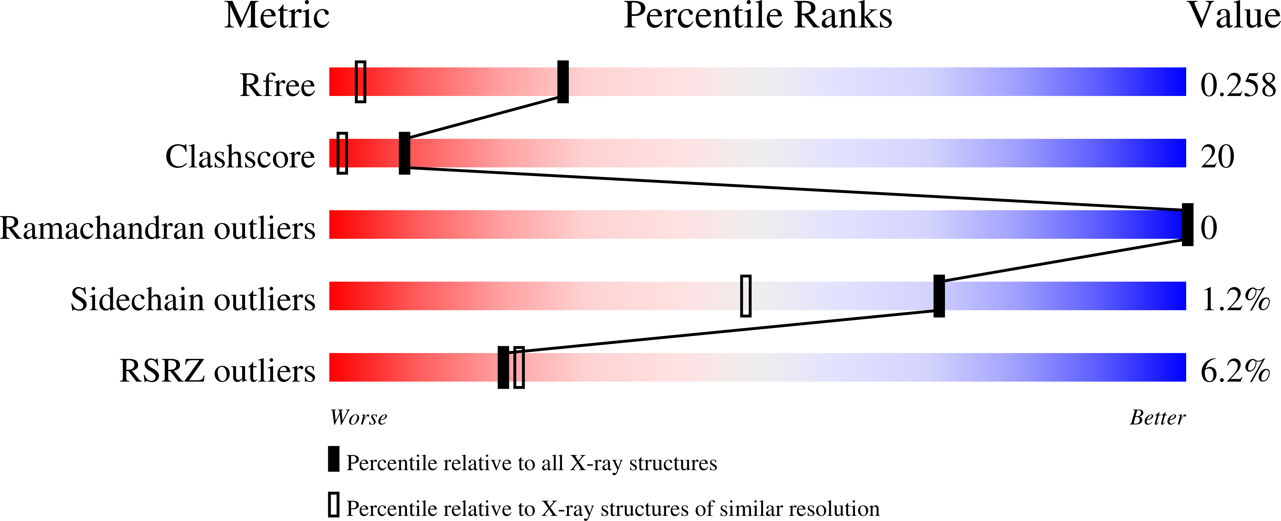
wwPDB Validation   3D Report Full Report


This is version 1.2 of the entry. See complete history


Literature

Three-Dimensional Structural Characterization of a Novel Drosophila Melanogaster Acylphosphatase

Zuccotti, S.Rosano, C.Ramazzotti, M.Degl'Innocenti, D.Stefani, M.Manao, G.Bolognesi, M.

(2004) Acta Crystallogr D Biol Crystallogr 60: 1177

  • DOI: https://doi.org/10.1107/S0907444904006808
  • Primary Citation of Related Structures:  
    1URR

  • PubMed Abstract: 

    Analysis of the Drosophila melanogaster EST database led to the discovery and cloning of a novel acylphosphatase. The CG18505 gene coding for a new enzyme (AcPDro2) is clearly distinct from the previously described CG16870Acyp gene, which also codes for a D. melanogaster acylphosphatase (AcPDro). The putative catalytic residues, together with residues held to stabilize the acylphosphatase fold, are conserved in the two encoded proteins. Crystals of AcPDro2, which belong to the trigonal space group P3(1)21, with unit-cell parameters a = b = 45.8, c = 98.6 angstroms, gamma = 120 degrees, allowed the solution of the protein structure by molecular replacement and its refinement to 1.5 angstroms resolution. The AcPDro2 active-site structure is discussed.


  • Organizational Affiliation

    Department of Physics-INFM and Center of Excellence for Biomedical Research, University of Genova, Via Dodecaneso 33, 16132 Genova, Italy.


Macromolecules
Find similar proteins by:  (by identity cutoff)  |  3D Structure
Entity ID: 1
MoleculeChains Sequence LengthOrganismDetailsImage
CG18505 PROTEIN102Drosophila melanogasterMutation(s): 0 
EC: 3.6.1.7
UniProt
Find proteins for Q9VF36 (Drosophila melanogaster)
Explore Q9VF36 
Go to UniProtKB:  Q9VF36
Entity Groups  
Sequence Clusters30% Identity50% Identity70% Identity90% Identity95% Identity100% Identity
UniProt GroupQ9VF36
Sequence Annotations
Expand
  • Reference Sequence
Small Molecules
Ligands 1 Unique
IDChains Name / Formula / InChI Key2D Diagram3D Interactions
GOL
Query on GOL

Download Ideal Coordinates CCD File 
B [auth A]GLYCEROL
C3 H8 O3
PEDCQBHIVMGVHV-UHFFFAOYSA-N
Experimental Data & Validation

Experimental Data

  • Method: X-RAY DIFFRACTION
  • Resolution: 1.50 Å
  • R-Value Free: 0.230 
  • R-Value Work: 0.160 
  • R-Value Observed: 0.163 
  • Space Group: P 31 2 1
Unit Cell:
Length ( Å )Angle ( ˚ )
a = 45.722α = 90
b = 45.722β = 90
c = 98.585γ = 120
Software Package:
Software NamePurpose
REFMACrefinement
MOSFLMdata reduction
SCALAdata scaling
AMoREphasing

Structure Validation

View Full Validation Report


Entry History 

Deposition Data

Revision History  (Full details and data files)

  • Version 1.0: 2004-05-27
    Type: Initial release
  • Version 1.1: 2014-02-19
    Changes: Database references, Derived calculations, Non-polymer description, Other, Structure summary, Version format compliance
  • Version 1.2: 2023-12-13
    Changes: Data collection, Database references, Derived calculations, Other, Refinement description