1Q4G

2.0 Angstrom Crystal Structure of Ovine Prostaglandin H2 Synthase-1, in complex with alpha-methyl-4-biphenylacetic acid


Experimental Data Snapshot

  • Method: X-RAY DIFFRACTION
  • Resolution: 2.00 Å
  • R-Value Free: 0.231 
  • R-Value Work: 0.217 
  • R-Value Observed: 0.217 

wwPDB Validation   3D Report Full Report


Ligand Structure Quality Assessment 


This is version 2.1 of the entry. See complete history


Literature

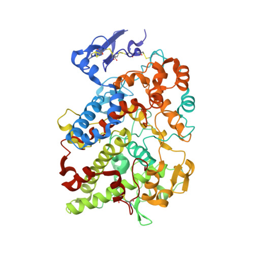
The 2.0A Resolution Crystal Structure of Prostaglandin H(2) Synthase-1: Structural Insights into an Unusual Peroxidase

Gupta, K.Selinsky, B.S.Kaub, C.J.Katz, A.K.Loll, P.J.

(2004) J Mol Biol 335: 503-518

  • DOI: https://doi.org/10.1016/j.jmb.2003.10.073
  • Primary Citation of Related Structures:  
    1Q4G

  • PubMed Abstract: 

    Prostaglandin H2 synthase (EC 1.14.99.1) is an integral membrane enzyme containing a cyclooxygenase site, which is the target for the non-steroidal anti-inflammatory drugs, and a spatially distinct peroxidase site. Previous crystallographic studies of this clinically important drug target have been hindered by low resolution. We present here the 2.0 A resolution X-ray crystal structure of ovine prostaglandin H2 synthase-1 in complex with alpha-methyl-4-biphenylacetic acid, a defluorinated analog of the non-steroidal anti-inflammatory drug flurbiprofen. Detergent molecules are seen to bind to the protein's membrane-binding domain, and their positions suggest the depth to which this domain is likely to penetrate into the lipid bilayer. The relation of the enzyme's proximal heme ligand His388 to the heme iron is atypical for a peroxidase; the iron-histidine bond is unusually long and a substantial tilt angle is observed between the heme and imidazole planes. A molecule of glycerol, used as a cryoprotectant during diffraction experiments, is seen to bind in the peroxidase site, offering the first view of any ligand in this active site. Insights gained from glycerol binding may prove useful in the design of a peroxidase-specific ligand.


  • Organizational Affiliation

    Department of Biochemistry, Drexel University College of Medicine, 245 N 15th Street, Mailstop 497, Philadelphia, PA 19102-1192, USA.


Macromolecules
Find similar proteins by:  (by identity cutoff)  |  3D Structure
Entity ID: 1
MoleculeChains Sequence LengthOrganismDetailsImage
Prostaglandin G/H synthase 1
A, B
553Ovis ariesMutation(s): 0 
EC: 1.14.99.1
Membrane Entity: Yes 
UniProt
Find proteins for P05979 (Ovis aries)
Explore P05979 
Go to UniProtKB:  P05979
Entity Groups  
Sequence Clusters30% Identity50% Identity70% Identity90% Identity95% Identity100% Identity
UniProt GroupP05979
Sequence Annotations
Expand
  • Reference Sequence
Oligosaccharides

Help

Entity ID: 2
MoleculeChains Length2D Diagram Glycosylation3D Interactions
2-acetamido-2-deoxy-alpha-D-glucopyranose-(1-4)-2-acetamido-2-deoxy-beta-D-glucopyranose
C, F
2N-Glycosylation
Glycosylation Resources
GlyTouCan:  G07375KG
GlyCosmos:  G07375KG
GlyGen:  G07375KG
Entity ID: 3
MoleculeChains Length2D Diagram Glycosylation3D Interactions
beta-D-mannopyranose-(1-6)-beta-D-mannopyranose-(1-4)-2-acetamido-2-deoxy-beta-D-glucopyranose-(1-4)-2-acetamido-2-deoxy-beta-D-glucopyranose
D, G
4N-Glycosylation
Glycosylation Resources
GlyTouCan:  G40894EI
GlyCosmos:  G40894EI
GlyGen:  G40894EI
Entity ID: 4
MoleculeChains Length2D Diagram Glycosylation3D Interactions
alpha-D-mannopyranose-(1-6)-alpha-D-mannopyranose-(1-6)-beta-D-mannopyranose-(1-4)-2-acetamido-2-deoxy-alpha-D-glucopyranose-(1-4)-2-acetamido-2-deoxy-beta-D-glucopyranose
E
5N-Glycosylation
Glycosylation Resources
GlyTouCan:  G42831WZ
GlyCosmos:  G42831WZ
GlyGen:  G42831WZ
Entity ID: 5
MoleculeChains Length2D Diagram Glycosylation3D Interactions
beta-D-mannopyranose-(1-6)-beta-D-mannopyranose-(1-6)-beta-D-mannopyranose-(1-4)-2-acetamido-2-deoxy-alpha-D-glucopyranose-(1-4)-2-acetamido-2-deoxy-beta-D-glucopyranose
H
5N-Glycosylation
Glycosylation Resources
GlyTouCan:  G07860ZO
GlyCosmos:  G07860ZO
GlyGen:  G07860ZO
Small Molecules
Ligands 4 Unique
IDChains Name / Formula / InChI Key2D Diagram3D Interactions
HEM
Query on HEM

Download Ideal Coordinates CCD File 
N [auth A],
V [auth B]
PROTOPORPHYRIN IX CONTAINING FE
C34 H32 Fe N4 O4
KABFMIBPWCXCRK-RGGAHWMASA-L
BOG
Query on BOG

Download Ideal Coordinates CCD File 
I [auth A]
J [auth A]
K [auth A]
L [auth A]
Q [auth B]
I [auth A],
J [auth A],
K [auth A],
L [auth A],
Q [auth B],
R [auth B],
S [auth B],
T [auth B]
octyl beta-D-glucopyranoside
C14 H28 O6
HEGSGKPQLMEBJL-RKQHYHRCSA-N
BFL
Query on BFL

Download Ideal Coordinates CCD File 
M [auth A],
U [auth B]
2-(1,1'-BIPHENYL-4-YL)PROPANOIC ACID
C15 H14 O2
JALUUBQFLPUJMY-NSHDSACASA-N
GOL
Query on GOL

Download Ideal Coordinates CCD File 
O [auth A],
P [auth A],
W [auth B],
X [auth B],
Y [auth B]
GLYCEROL
C3 H8 O3
PEDCQBHIVMGVHV-UHFFFAOYSA-N
Binding Affinity Annotations 
IDSourceBinding Affinity
BFL BindingDB:  1Q4G Ki: 130 (nM) from 1 assay(s)
IC50: min: 10, max: 110 (nM) from 3 assay(s)
Experimental Data & Validation

Experimental Data

  • Method: X-RAY DIFFRACTION
  • Resolution: 2.00 Å
  • R-Value Free: 0.231 
  • R-Value Work: 0.217 
  • R-Value Observed: 0.217 
  • Space Group: I 2 2 2
Unit Cell:
Length ( Å )Angle ( ˚ )
a = 98.147α = 90
b = 203.859β = 90
c = 223.599γ = 90
Software Package:
Software NamePurpose
CNSrefinement
CNSphasing

Structure Validation

View Full Validation Report


Ligand Structure Quality Assessment 


Entry History 

Deposition Data

Revision History  (Full details and data files)

  • Version 1.0: 2004-01-06
    Type: Initial release
  • Version 1.1: 2008-04-29
    Changes: Version format compliance
  • Version 1.2: 2011-07-13
    Changes: Non-polymer description, Version format compliance
  • Version 1.3: 2014-11-12
    Changes: Derived calculations
  • Version 2.0: 2020-07-29
    Type: Remediation
    Reason: Carbohydrate remediation
    Changes: Atomic model, Data collection, Derived calculations, Structure summary
  • Version 2.1: 2023-08-16
    Changes: Data collection, Database references, Refinement description, Structure summary