1PC8

Crystal Structure of a novel form of mistletoe lectin from Himalayan Viscum album L. at 3.8A resolution


Experimental Data Snapshot

  • Method: X-RAY DIFFRACTION
  • Resolution: 3.80 Å
  • R-Value Free: 0.271 
  • R-Value Work: 0.228 
  • R-Value Observed: 0.231 

wwPDB Validation   3D Report Full Report


Ligand Structure Quality Assessment 


This is version 2.1 of the entry. See complete history


Literature

Structure of a novel ribosome-inactivating protein from a hemi-parasitic plant inhabiting the northwestern Himalayas.

Mishra, V.Ethayathulla, A.S.Sharma, R.S.Yadav, S.Krauspenhaar, R.Betzel, C.Babu, C.R.Singh, T.P.

(2004) Acta Crystallogr D Biol Crystallogr 60: 2295-2304

  • DOI: https://doi.org/10.1107/S0907444904023534
  • Primary Citation of Related Structures:  
    1PC8

  • PubMed Abstract: 

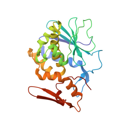
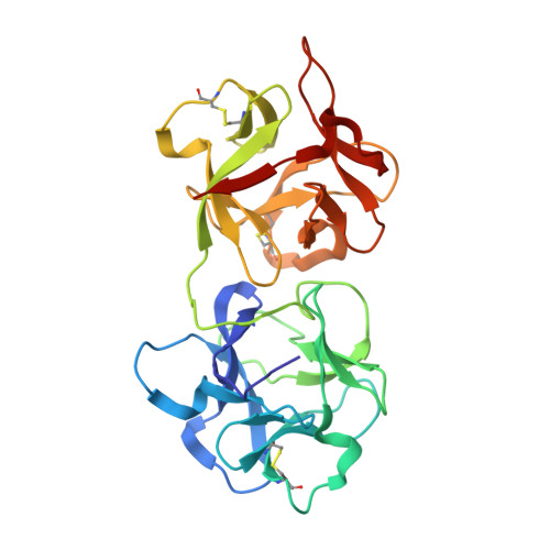
    This is the first report of the structural studies of a novel ribosome-inactivating protein (RIP) obtained from the Himalayan mistletoe (Viscum album) (HmRip). HmRip is a type II heterodimeric protein consisting of a toxic enzyme (A-chain) with an active site for ribosome inactivation and a lectin subunit (B-chain) with well defined sugar-binding sites. The crystal structure of HmRip has been determined at 3.8 A resolution and refined to a crystallographic R factor of 0.228 (R(free) = 0.271). A comparison of this structure with other type II RIPs reveals the presence of distinct structural features in the active site of the A-chain and in the 2gamma sugar-binding site of the B-chain. The conformation of the side chain of Tyr110, which is a conserved active-site residue in the A subunit, is strikingly different from those observed in other mistletoe RIPs, indicating its unique substrate-binding preference. The deletion of two important residues from the kink region after Ala231 in the 2gamma subdomain of the B-chain results in a significantly different conformation of the sugar-binding pocket. A ribosome-recognition site has also been identified in HmRip. The site is a shallow cavity, with the conserved residues Arg51, Asp70, Thr72 and Asn73 involved in the binding. The conformations of the antigenic epitopes of residues 1-20, 85-103 and 206-223 differ from those observed in other type II RIPs, resulting in the distinct antigenicity and pharmacological properties of HmRip.


  • Organizational Affiliation

    Department of Biophysics, All India Institute of Medical Sciences, New Delhi, India.


Macromolecules
Find similar proteins by:  (by identity cutoff)  |  3D Structure
Entity ID: 1
MoleculeChains Sequence LengthOrganismDetailsImage
Himalayan mistletoe ribosome-inactivating protein240Viscum albumMutation(s): 0 
EC: 3.2.2.22
UniProt
Find proteins for Q6ITZ3 (Viscum album)
Explore Q6ITZ3 
Go to UniProtKB:  Q6ITZ3
Entity Groups  
Sequence Clusters30% Identity50% Identity70% Identity90% Identity95% Identity100% Identity
UniProt GroupQ6ITZ3
Sequence Annotations
Expand
  • Reference Sequence
Find similar proteins by:  (by identity cutoff)  |  3D Structure
Entity ID: 2
MoleculeChains Sequence LengthOrganismDetailsImage
Himalayan mistletoe ribosome-inactivating protein255Viscum albumMutation(s): 0 
EC: 3.2.2.22
UniProt
Find proteins for Q6ITZ3 (Viscum album)
Explore Q6ITZ3 
Go to UniProtKB:  Q6ITZ3
Entity Groups  
Sequence Clusters30% Identity50% Identity70% Identity90% Identity95% Identity100% Identity
UniProt GroupQ6ITZ3
Sequence Annotations
Expand
  • Reference Sequence
Oligosaccharides

Help

Entity ID: 3
MoleculeChains Length2D Diagram Glycosylation3D Interactions
2-acetamido-2-deoxy-beta-D-glucopyranose-(1-4)-2-acetamido-2-deoxy-beta-D-glucopyranose
C, E
2N-Glycosylation
Glycosylation Resources
GlyTouCan:  G42666HT
GlyCosmos:  G42666HT
GlyGen:  G42666HT
Entity ID: 4
MoleculeChains Length2D Diagram Glycosylation3D Interactions
beta-D-mannopyranose-(1-4)-2-acetamido-2-deoxy-beta-D-glucopyranose-(1-4)-2-acetamido-2-deoxy-beta-D-glucopyranose
D
3N-Glycosylation
Glycosylation Resources
GlyTouCan:  G15407YE
GlyCosmos:  G15407YE
GlyGen:  G15407YE
Small Molecules
Ligands 1 Unique
IDChains Name / Formula / InChI Key2D Diagram3D Interactions
NAG
Query on NAG

Download Ideal Coordinates CCD File 
F [auth A]2-acetamido-2-deoxy-beta-D-glucopyranose
C8 H15 N O6
OVRNDRQMDRJTHS-FMDGEEDCSA-N
Experimental Data & Validation

Experimental Data

  • Method: X-RAY DIFFRACTION
  • Resolution: 3.80 Å
  • R-Value Free: 0.271 
  • R-Value Work: 0.228 
  • R-Value Observed: 0.231 
  • Space Group: P 65 2 2
Unit Cell:
Length ( Å )Angle ( ˚ )
a = 109.182α = 90
b = 109.182β = 90
c = 309.44γ = 120
Software Package:
Software NamePurpose
REFMACrefinement
MAR345data collection
SCALEPACKdata scaling
AMoREphasing

Structure Validation

View Full Validation Report


Ligand Structure Quality Assessment 


Entry History 

Deposition Data

Revision History  (Full details and data files)

  • Version 1.0: 2004-06-22
    Type: Initial release
  • Version 1.1: 2008-04-29
    Changes: Version format compliance
  • Version 1.2: 2011-07-13
    Changes: Non-polymer description, Version format compliance
  • Version 1.3: 2017-10-11
    Changes: Refinement description
  • Version 2.0: 2020-07-29
    Type: Remediation
    Reason: Carbohydrate remediation
    Changes: Advisory, Atomic model, Data collection, Derived calculations, Non-polymer description, Structure summary
  • Version 2.1: 2023-10-25
    Changes: Data collection, Database references, Refinement description, Structure summary