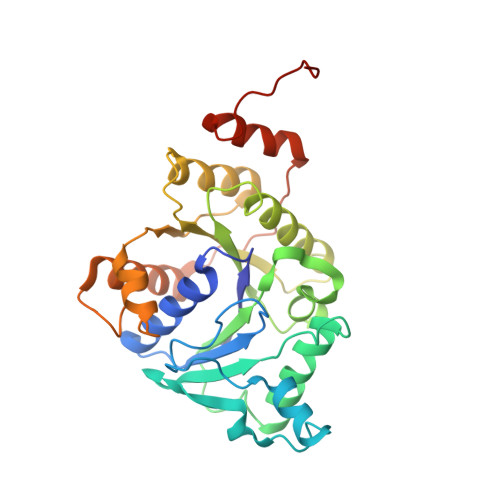
Crystallographic structure of the nitrogenase iron protein from Azotobacter vinelandii.
Georgiadis, M.M., Komiya, H., Chakrabarti, P., Woo, D., Kornuc, J.J., Rees, D.C.(1992) Science 257: 1653-1659
- PubMed: 1529353
- DOI: https://doi.org/10.1126/science.1529353
- Primary Citation of Related Structures:
1NIP - PubMed Abstract:
The nitrogenase enzyme system catalyzes the ATP (adenosine triphosphate)-dependent reduction of dinitrogen to ammonia during the process of nitrogen fixation. Nitrogenase consists of two proteins: the iron (Fe)-protein, which couples hydrolysis of ATP to electron transfer, and the molybdenum-iron (MoFe)-protein, which contains the dinitrogen binding site. In order to address the role of ATP in nitrogen fixation, the crystal structure of the nitrogenase Fe-protein from Azotobacter vinelandii has been determined at 2.9 angstrom (A) resolution. Fe-protein is a dimer of two identical subunits that coordinate a single 4Fe:4S cluster. Each subunit folds as a single alpha/beta type domain, which together symmetrically ligate the surface exposed 4Fe:4S cluster through two cysteines from each subunit. A single bound ADP (adenosine diphosphate) molecule is located in the interface region between the two subunits. Because the phosphate groups of this nucleotide are approximately 20 A from the 4Fe:4S cluster, it is unlikely that ATP hydrolysis and electron transfer are directly coupled. Instead, it appears that interactions between the nucleotide and cluster sites must be indirectly coupled by allosteric changes occurring at the subunit interface. The coupling between protein conformation and nucleotide hydrolysis in Fe-protein exhibits general similarities to the H-Ras p21 and recA proteins that have been recently characterized structurally. The Fe-protein structure may be relevant to the functioning of other biochemical energy-transducing systems containing two nucleotide-binding sites, including membrane transport proteins.
Organizational Affiliation:
Department of Biochemistry, Columbia University, New York, NY 10032.