1MYR

MYROSINASE FROM SINAPIS ALBA


Experimental Data Snapshot

  • Method: X-RAY DIFFRACTION
  • Resolution: 1.64 Å
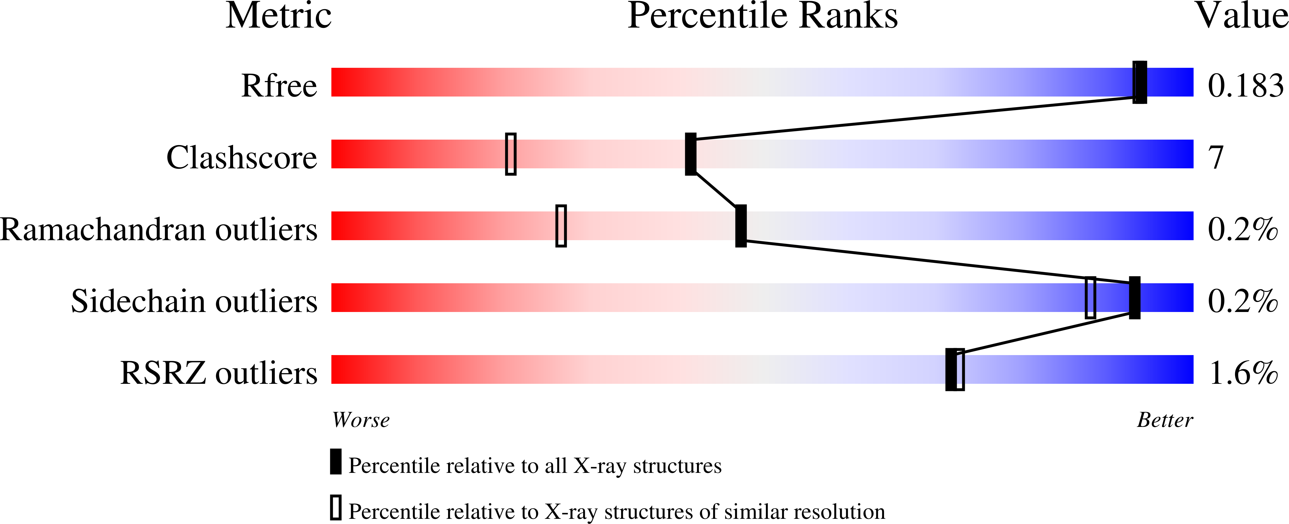
  • R-Value Free: 0.187 
  • R-Value Work: 0.152 
  • R-Value Observed: 0.152 

wwPDB Validation   3D Report Full Report


Ligand Structure Quality Assessment 


This is version 2.1 of the entry. See complete history


Literature

The crystal structures of Sinapis alba myrosinase and a covalent glycosyl-enzyme intermediate provide insights into the substrate recognition and active-site machinery of an S-glycosidase.

Burmeister, W.P.Cottaz, S.Driguez, H.Iori, R.Palmieri, S.Henrissat, B.

(1997) Structure 5: 663-675

  • DOI: https://doi.org/10.1016/s0969-2126(97)00221-9
  • Primary Citation of Related Structures:  
    1MYR

  • PubMed Abstract: 

    Myrosinase is the enzyme responsible for the hydrolysis of a variety of plant anionic 1-thio-beta-D-glucosides called glucosinolates. Myrosinase and glucosinolates, which are stored in different tissues of the plant, are mixed during mastication generating toxic by-products that are believed to play a role in the plant defence system. Whilst O-glycosidases are extremely widespread in nature, myrosinase is the only known S-glycosidase. This intriguing enzyme, which shows sequence similarities with O-glycosidases, offers the opportunity to analyze the similarities and differences between enzymes hydrolyzing S- and O-glycosidic bonds.


  • Organizational Affiliation

    European Synchrotron Radiation Facility (ESRF), BP 220, F-38043 Grenoble cedex, France. wpb@esrf.fr


Macromolecules
Find similar proteins by:  (by identity cutoff)  |  3D Structure
Entity ID: 1
MoleculeChains Sequence LengthOrganismDetailsImage
MYROSINASE501Sinapis albaMutation(s): 0 
EC: 3.2.3.1
UniProt
Find proteins for P29736 (Sinapis alba)
Explore P29736 
Go to UniProtKB:  P29736
Entity Groups  
Sequence Clusters30% Identity50% Identity70% Identity90% Identity95% Identity100% Identity
UniProt GroupP29736
Sequence Annotations
Expand
  • Reference Sequence
Oligosaccharides

Help

Entity ID: 2
MoleculeChains Length2D Diagram Glycosylation3D Interactions
2-acetamido-2-deoxy-beta-D-glucopyranose-(1-4)-2-acetamido-2-deoxy-beta-D-glucopyranose
B, E
2N-Glycosylation
Glycosylation Resources
GlyTouCan:  G42666HT
GlyCosmos:  G42666HT
GlyGen:  G42666HT
Entity ID: 3
MoleculeChains Length2D Diagram Glycosylation3D Interactions
beta-D-xylopyranose-(1-2)-beta-D-mannopyranose-(1-4)-2-acetamido-2-deoxy-beta-D-glucopyranose-(1-4)-[alpha-L-fucopyranose-(1-3)]2-acetamido-2-deoxy-beta-D-glucopyranose
C
5N-Glycosylation
Glycosylation Resources
GlyTouCan:  G06206UV
GlyCosmos:  G06206UV
GlyGen:  G06206UV
Entity ID: 4
MoleculeChains Length2D Diagram Glycosylation3D Interactions
beta-D-xylopyranose-(1-2)-[alpha-D-mannopyranose-(1-3)][alpha-D-mannopyranose-(1-6)]beta-D-mannopyranose-(1-4)-2-acetamido-2-deoxy-beta-D-glucopyranose-(1-4)-[alpha-L-fucopyranose-(1-3)]2-acetamido-2-deoxy-beta-D-glucopyranose
D
7N-Glycosylation
Glycosylation Resources
GlyTouCan:  G26295XE
GlyCosmos:  G26295XE
GlyGen:  G26295XE
Small Molecules
Ligands 4 Unique
IDChains Name / Formula / InChI Key2D Diagram3D Interactions
NAG
Query on NAG

Download Ideal Coordinates CCD File 
F [auth A],
G [auth A],
H [auth A],
I [auth A],
J [auth A]
2-acetamido-2-deoxy-beta-D-glucopyranose
C8 H15 N O6
OVRNDRQMDRJTHS-FMDGEEDCSA-N
SO4
Query on SO4

Download Ideal Coordinates CCD File 
L [auth A]
M [auth A]
N [auth A]
O [auth A]
P [auth A]
L [auth A],
M [auth A],
N [auth A],
O [auth A],
P [auth A],
Q [auth A],
R [auth A],
S [auth A],
T [auth A],
U [auth A]
SULFATE ION
O4 S
QAOWNCQODCNURD-UHFFFAOYSA-L
GOL
Query on GOL

Download Ideal Coordinates CCD File 
V [auth A],
W [auth A],
X [auth A],
Y [auth A]
GLYCEROL
C3 H8 O3
PEDCQBHIVMGVHV-UHFFFAOYSA-N
ZN
Query on ZN

Download Ideal Coordinates CCD File 
K [auth A]ZINC ION
Zn
PTFCDOFLOPIGGS-UHFFFAOYSA-N
Experimental Data & Validation

Experimental Data

  • Method: X-RAY DIFFRACTION
  • Resolution: 1.64 Å
  • R-Value Free: 0.187 
  • R-Value Work: 0.152 
  • R-Value Observed: 0.152 
  • Space Group: C 2 2 21
Unit Cell:
Length ( Å )Angle ( ˚ )
a = 134.3α = 90
b = 136.4β = 90
c = 80.3γ = 90
Software Package:
Software NamePurpose
MOSFLMdata reduction
ROTAVATAdata reduction
Agrovatadata reduction
X-PLORmodel building
X-PLORrefinement
CCP4data scaling
ROTAVATAdata scaling
X-PLORphasing

Structure Validation

View Full Validation Report


Ligand Structure Quality Assessment 


Entry History 

Deposition Data

Revision History  (Full details and data files)

  • Version 1.0: 1997-06-16
    Type: Initial release
  • Version 1.1: 2008-03-24
    Changes: Version format compliance
  • Version 1.2: 2011-07-13
    Changes: Derived calculations, Version format compliance
  • Version 1.3: 2011-11-16
    Changes: Atomic model
  • Version 2.0: 2020-07-29
    Type: Remediation
    Reason: Carbohydrate remediation
    Changes: Advisory, Atomic model, Data collection, Derived calculations, Non-polymer description, Refinement description, Structure summary
  • Version 2.1: 2023-08-09
    Changes: Database references, Derived calculations, Refinement description, Structure summary