1I3H

CONCANAVALIN A-DIMANNOSE STRUCTURE


Experimental Data Snapshot

  • Method: X-RAY DIFFRACTION
  • Resolution: 1.20 Å
  • R-Value Free: 0.190 
  • R-Value Work: 0.167 
  • R-Value Observed: 0.170 

wwPDB Validation   3D Report Full Report


This is version 2.1 of the entry. See complete history


Literature

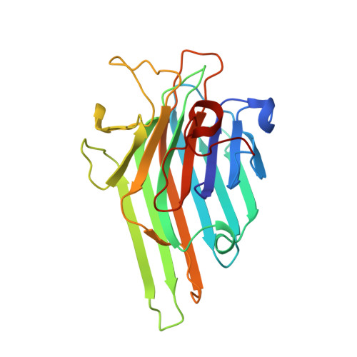
The 1.2 A resolution structure of the Con A-dimannose complex.

Sanders, D.A.Moothoo, D.N.Raftery, J.Howard, A.J.Helliwell, J.R.Naismith, J.H.

(2001) J Mol Biol 310: 875-884

  • DOI: https://doi.org/10.1006/jmbi.2001.4806
  • Primary Citation of Related Structures:  
    1I3H

  • PubMed Abstract: 

    The complex between concanavalin A (Con A) and alpha1-2 mannobiose (mannose alpha1-2 mannose) has been refined to 1.2 A resolution. This is the highest resolution structure reported for any sugar-lectin complex. As the native structure of Con A to 0.94 A resolution is already in the database, this gives us a unique opportunity to examine sugar-protein binding at high resolution. These data have allowed us to model a number of hydrogen atoms involved in the binding of the sugar to Con A, using the difference density map to place the hydrogen atoms. This map reveals the presence of the protonated form of Asp208 involved in binding. Asp208 is not protonated in the 0.94 A native structure. Our results clearly show that this residue is protonated and hydrogen bonds to the sugar. The structure accounts for the higher affinity of the alpha1-2 linked sugar when compared to other disaccharides. This structure identifies different interactions to those predicted by previous modelling studies. We believe that the additional data presented here will enable significant improvements to be made to the sugar-protein modelling algorithms.


  • Organizational Affiliation

    Biomolecular Sciences, The University, St. Andrews, KY16 9ST, Scotland.


Macromolecules
Find similar proteins by:  (by identity cutoff)  |  3D Structure
Entity ID: 1
MoleculeChains Sequence LengthOrganismDetailsImage
Concanavalin-A237Canavalia ensiformisMutation(s): 0 
UniProt
Find proteins for P02866 (Canavalia ensiformis)
Explore P02866 
Go to UniProtKB:  P02866
Entity Groups  
Sequence Clusters30% Identity50% Identity70% Identity90% Identity95% Identity100% Identity
UniProt GroupP02866
Sequence Annotations
Expand
  • Reference Sequence
Oligosaccharides

Help

Entity ID: 2
MoleculeChains Length2D Diagram Glycosylation3D Interactions
alpha-D-mannopyranose-(1-2)-alpha-D-mannopyranose
B
2N/A
Glycosylation Resources
GlyTouCan:  G53402KW
GlyCosmos:  G53402KW
GlyGen:  G53402KW
Small Molecules
Biologically Interesting Molecules (External Reference) 1 Unique
Experimental Data & Validation

Experimental Data

  • Method: X-RAY DIFFRACTION
  • Resolution: 1.20 Å
  • R-Value Free: 0.190 
  • R-Value Work: 0.167 
  • R-Value Observed: 0.170 
  • Space Group: I 2 2 2
Unit Cell:
Length ( Å )Angle ( ˚ )
a = 90.9α = 90
b = 86.41β = 90
c = 65.42γ = 90
Software Package:
Software NamePurpose
AMoREphasing
REFMACrefinement
DENZOdata reduction
CCP4data scaling

Structure Validation

View Full Validation Report


Entry History 

Deposition Data

Revision History  (Full details and data files)

  • Version 1.0: 2001-07-25
    Type: Initial release
  • Version 1.1: 2008-04-27
    Changes: Version format compliance
  • Version 1.2: 2011-07-13
    Changes: Derived calculations, Version format compliance
  • Version 1.3: 2017-06-28
    Changes: Data collection, Database references, Source and taxonomy, Structure summary
  • Version 2.0: 2020-07-29
    Type: Remediation
    Reason: Carbohydrate remediation
    Changes: Atomic model, Data collection, Derived calculations, Structure summary
  • Version 2.1: 2024-02-07
    Changes: Advisory, Data collection, Database references, Structure summary