1FMW

CRYSTAL STRUCTURE OF THE MGATP COMPLEX FOR THE MOTOR DOMAIN OF DICTYOSTELIUM MYOSIN II


Experimental Data Snapshot

  • Method: X-RAY DIFFRACTION
  • Resolution: 2.15 Å
  • R-Value Free: 0.286 
  • R-Value Work: 0.206 
  • R-Value Observed: 0.206 

wwPDB Validation   3D Report Full Report


Ligand Structure Quality Assessment 


This is version 1.4 of the entry. See complete history


Literature

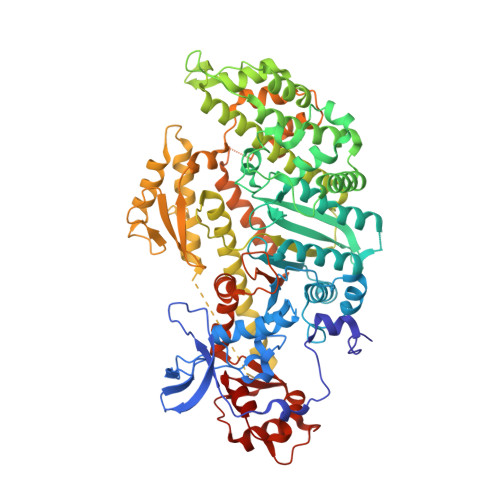
X-ray structures of the apo and MgATP-bound states of Dictyostelium discoideum myosin motor domain.

Bauer, C.B.Holden, H.M.Thoden, J.B.Smith, R.Rayment, I.

(2000) J Biol Chem 275: 38494-38499

  • DOI: https://doi.org/10.1074/jbc.M005585200
  • Primary Citation of Related Structures:  
    1FMV, 1FMW

  • PubMed Abstract: 

    Myosin is the most comprehensively studied molecular motor that converts energy from the hydrolysis of MgATP into directed movement. Its motile cycle consists of a sequential series of interactions between myosin, actin, MgATP, and the products of hydrolysis, where the affinity of myosin for actin is modulated by the nature of the nucleotide bound in the active site. The first step in the contractile cycle occurs when ATP binds to actomyosin and releases myosin from the complex. We report here the structure of the motor domain of Dictyostelium discoideum myosin II both in its nucleotide-free state and complexed with MgATP. The structure with MgATP was obtained by soaking the crystals in substrate. These structures reveal that both the apo form and the MgATP complex are very similar to those previously seen with MgATPgammaS and MgAMP-PNP. Moreover, these structures are similar to that of chicken skeletal myosin subfragment-1. The crystallized protein is enzymatically active in solution, indicating that the conformation of myosin observed in chicken skeletal myosin subfragment-1 is unable to hydrolyze ATP and most likely represents the pre-hydrolysis structure for the myosin head that occurs after release from actin.


  • Organizational Affiliation

    Department of Biochemistry, University of Wisconsin, Madison, Wisconsin 53706-1544, USA.


Macromolecules
Find similar proteins by:  (by identity cutoff)  |  3D Structure
Entity ID: 1
MoleculeChains Sequence LengthOrganismDetailsImage
MYOSIN II HEAVY CHAIN761Dictyostelium discoideumMutation(s): 0 
UniProt
Find proteins for P08799 (Dictyostelium discoideum)
Explore P08799 
Go to UniProtKB:  P08799
Entity Groups  
Sequence Clusters30% Identity50% Identity70% Identity90% Identity95% Identity100% Identity
UniProt GroupP08799
Sequence Annotations
Expand
  • Reference Sequence
Small Molecules
Ligands 2 Unique
IDChains Name / Formula / InChI Key2D Diagram3D Interactions
ATP
Query on ATP

Download Ideal Coordinates CCD File 
C [auth A]ADENOSINE-5'-TRIPHOSPHATE
C10 H16 N5 O13 P3
ZKHQWZAMYRWXGA-KQYNXXCUSA-N
MG
Query on MG

Download Ideal Coordinates CCD File 
B [auth A]MAGNESIUM ION
Mg
JLVVSXFLKOJNIY-UHFFFAOYSA-N
Experimental Data & Validation

Experimental Data

  • Method: X-RAY DIFFRACTION
  • Resolution: 2.15 Å
  • R-Value Free: 0.286 
  • R-Value Work: 0.206 
  • R-Value Observed: 0.206 
  • Space Group: P 21 21 2
Unit Cell:
Length ( Å )Angle ( ˚ )
a = 104.1α = 90
b = 180.4β = 90
c = 54.2γ = 90
Software Package:
Software NamePurpose
AMoREphasing
TNTrefinement
DENZOdata reduction
CCP4data scaling

Structure Validation

View Full Validation Report


Ligand Structure Quality Assessment 


Entry History 

Deposition Data

Revision History  (Full details and data files)

  • Version 1.0: 2000-11-22
    Type: Initial release
  • Version 1.1: 2008-04-27
    Changes: Version format compliance
  • Version 1.2: 2011-07-13
    Changes: Version format compliance
  • Version 1.3: 2012-11-21
    Changes: Atomic model, Experimental preparation
  • Version 1.4: 2024-02-07
    Changes: Data collection, Database references, Derived calculations