1F2Q

CRYSTAL STRUCTURE OF THE HUMAN HIGH-AFFINITY IGE RECEPTOR


Experimental Data Snapshot

  • Method: X-RAY DIFFRACTION
  • Resolution: 2.40 Å
  • R-Value Free: 0.254 
  • R-Value Work: 0.226 
  • R-Value Observed: 0.226 

wwPDB Validation   3D Report Full Report


This is version 2.0 of the entry. See complete history


Literature

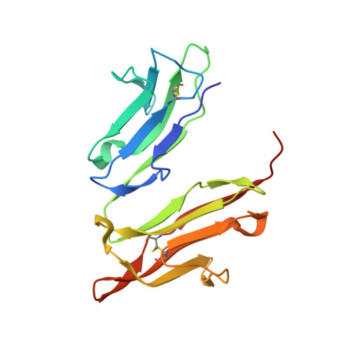
Crystal structure of the human high-affinity IgE receptor.

Garman, S.C.Kinet, J.P.Jardetzky, T.S.

(1998) Cell 95: 951-961

  • DOI: https://doi.org/10.1016/s0092-8674(00)81719-5
  • Primary Citation of Related Structures:  
    1F2Q

  • PubMed Abstract: 

    Allergic responses result from the activation of mast cells by the human high-affinity IgE receptor. IgE-mediated allergic reactions may develop to a variety of environmental compounds, but the initiation of a response requires the binding of IgE to its high-affinity receptor. We have solved the X-ray crystal structure of the antibody-binding domains of the human IgE receptor at 2.4 A resolution. The structure reveals a highly bent arrangement of immunoglobulin domains that form an extended convex surface of interaction with IgE. A prominent loop that confers specificity for IgE molecules extends from the receptor surface near an unusual arrangement of four exposed tryptophans. The crystal structure of the IgE receptor provides a foundation for the development of new therapeutic approaches to allergy treatment.


  • Organizational Affiliation

    Department of Biochemistry, Molecular Biology, and Cell Biology, Northwestern University, Evanston, Illinois 60208, USA.


Macromolecules
Find similar proteins by:  (by identity cutoff)  |  3D Structure
Entity ID: 1
MoleculeChains Sequence LengthOrganismDetailsImage
HIGH AFFINITY IMMUNOGLOBULIN EPSILON RECEPTOR ALPHA-SUBUNIT176Homo sapiensMutation(s): 0 
UniProt & NIH Common Fund Data Resources
Find proteins for P12319 (Homo sapiens)
Explore P12319 
Go to UniProtKB:  P12319
PHAROS:  P12319
GTEx:  ENSG00000179639 
Entity Groups  
Sequence Clusters30% Identity50% Identity70% Identity90% Identity95% Identity100% Identity
UniProt GroupP12319
Sequence Annotations
Expand
  • Reference Sequence
Oligosaccharides

Help

Entity ID: 2
MoleculeChains Length2D Diagram Glycosylation3D Interactions
alpha-D-mannopyranose-(1-4)-2-acetamido-2-deoxy-beta-D-glucopyranose-(1-4)-2-acetamido-2-deoxy-beta-D-glucopyranose
B, C
3N-Glycosylation
Glycosylation Resources
GlyTouCan:  G62182OO
GlyCosmos:  G62182OO
GlyGen:  G62182OO
Small Molecules
Ligands 1 Unique
IDChains Name / Formula / InChI Key2D Diagram3D Interactions
NAG
Query on NAG

Download Ideal Coordinates CCD File 
D [auth A]2-acetamido-2-deoxy-beta-D-glucopyranose
C8 H15 N O6
OVRNDRQMDRJTHS-FMDGEEDCSA-N
Experimental Data & Validation

Experimental Data

  • Method: X-RAY DIFFRACTION
  • Resolution: 2.40 Å
  • R-Value Free: 0.254 
  • R-Value Work: 0.226 
  • R-Value Observed: 0.226 
  • Space Group: C 1 2 1
Unit Cell:
Length ( Å )Angle ( ˚ )
a = 88.59α = 90
b = 69.64β = 116.69
c = 49.36γ = 90
Software Package:
Software NamePurpose
DENZOdata reduction
SCALEPACKdata scaling
DMmodel building
CNSrefinement
DMphasing

Structure Validation

View Full Validation Report


Entry History 

Deposition Data

Revision History  (Full details and data files)

  • Version 1.0: 2000-06-08
    Type: Initial release
  • Version 1.1: 2008-04-27
    Changes: Version format compliance
  • Version 1.2: 2011-07-13
    Changes: Non-polymer description, Version format compliance
  • Version 2.0: 2020-07-29
    Type: Remediation
    Reason: Carbohydrate remediation
    Changes: Advisory, Atomic model, Data collection, Derived calculations, Structure summary