1BCU

ALPHA-THROMBIN COMPLEXED WITH HIRUGEN AND PROFLAVIN


Experimental Data Snapshot

  • Method: X-RAY DIFFRACTION
  • Resolution: 2.00 Å
  • R-Value Free: 0.212 
  • R-Value Work: 0.189 
  • R-Value Observed: 0.189 

wwPDB Validation   3D Report Full Report


Ligand Structure Quality Assessment 


This is version 1.4 of the entry. See complete history


Literature

X-ray and spectrophotometric studies of the binding of proflavin to the S1 specificity pocket of human alpha-thrombin.

Conti, E.Rivetti, C.Wonacott, A.Brick, P.

(1998) FEBS Lett 425: 229-233

  • DOI: https://doi.org/10.1016/s0014-5793(98)00235-x
  • Primary Citation of Related Structures:  
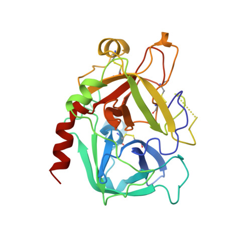
    1BCU

  • PubMed Abstract: 

    Proflavin can be used to study the interactions of inhibitors and substrates with thrombin by monitoring the changes in the visible absorption spectrum that occur on dye displacement. We have used microspectrophotometric methods to investigate the binding of proflavin to crystals of an alpha-thrombin-hirugen complex and have determined the structure by X-ray crystallography. The proflavin molecule binds in the S1 pocket of the enzyme with one of the amino groups hydrogen bonded to the carboxylate of Asp-189 while the protonated ring nitrogen is hydrogen bonded to the carbonyl of Gly-219. This result indicates that the proflavin displacement assay can be used to specifically monitor the binding of inhibitors to the S1 pocket.


  • Organizational Affiliation

    Blackett Laboratory, Imperial College, London, UK.


Macromolecules
Find similar proteins by:  (by identity cutoff)  |  3D Structure
Entity ID: 1
MoleculeChains Sequence LengthOrganismDetailsImage
ALPHA-THROMBINA [auth L]36Homo sapiensMutation(s): 0 
EC: 3.4.21.5
UniProt & NIH Common Fund Data Resources
Find proteins for P00734 (Homo sapiens)
Explore P00734 
Go to UniProtKB:  P00734
PHAROS:  P00734
GTEx:  ENSG00000180210 
Entity Groups  
Sequence Clusters30% Identity50% Identity70% Identity90% Identity95% Identity100% Identity
UniProt GroupP00734
Sequence Annotations
Expand
  • Reference Sequence
Find similar proteins by:  (by identity cutoff)  |  3D Structure
Entity ID: 2
MoleculeChains Sequence LengthOrganismDetailsImage
ALPHA-THROMBINB [auth H]259Homo sapiensMutation(s): 0 
EC: 3.4.21.5
UniProt & NIH Common Fund Data Resources
Find proteins for P00734 (Homo sapiens)
Explore P00734 
Go to UniProtKB:  P00734
PHAROS:  P00734
GTEx:  ENSG00000180210 
Entity Groups  
Sequence Clusters30% Identity50% Identity70% Identity90% Identity95% Identity100% Identity
UniProt GroupP00734
Sequence Annotations
Expand
  • Reference Sequence

Find similar proteins by:  Sequence   |   3D Structure  

Entity ID: 3
MoleculeChains Sequence LengthOrganismDetailsImage
HIRUGENC [auth I]12Hirudo medicinalisMutation(s): 0 
UniProt
Find proteins for P01050 (Hirudo medicinalis)
Explore P01050 
Go to UniProtKB:  P01050
Entity Groups  
Sequence Clusters30% Identity50% Identity70% Identity90% Identity95% Identity100% Identity
UniProt GroupP01050
Sequence Annotations
Expand
  • Reference Sequence
Small Molecules
Ligands 1 Unique
IDChains Name / Formula / InChI Key2D Diagram3D Interactions
PRL
Query on PRL

Download Ideal Coordinates CCD File 
D [auth H]PROFLAVIN
C13 H11 N3
WDVSHHCDHLJJJR-UHFFFAOYSA-N
Modified Residues  1 Unique
IDChains TypeFormula2D DiagramParent
TYS
Query on TYS
C [auth I]L-PEPTIDE LINKINGC9 H11 N O6 STYR
Binding Affinity Annotations 
IDSourceBinding Affinity
PRL PDBBind:  1BCU Kd: 5.30e+5 (nM) from 1 assay(s)
Binding MOAD:  1BCU Kd: 5.30e+5 (nM) from 1 assay(s)
Experimental Data & Validation

Experimental Data

  • Method: X-RAY DIFFRACTION
  • Resolution: 2.00 Å
  • R-Value Free: 0.212 
  • R-Value Work: 0.189 
  • R-Value Observed: 0.189 
  • Space Group: C 1 2 1
Unit Cell:
Length ( Å )Angle ( ˚ )
a = 71.15α = 90
b = 72.25β = 100.61
c = 72.67γ = 90
Software Package:
Software NamePurpose
MADNESdata collection
CCP4data reduction
X-PLORmodel building
X-PLORrefinement
MADNESdata reduction
CCP4data scaling
X-PLORphasing

Structure Validation

View Full Validation Report


Ligand Structure Quality Assessment 


Entry History 

Deposition Data

Revision History  (Full details and data files)

  • Version 1.0: 1998-10-14
    Type: Initial release
  • Version 1.1: 2008-03-03
    Changes: Version format compliance
  • Version 1.2: 2011-07-13
    Changes: Atomic model, Database references, Derived calculations, Non-polymer description, Structure summary, Version format compliance
  • Version 1.3: 2018-04-18
    Changes: Data collection, Other
  • Version 1.4: 2023-08-02
    Changes: Database references, Derived calculations, Refinement description