9WCW | pdb_00009wcw

AMP-PNP bound E.coli CnoX-GroEL complex, state I


Experimental Data Snapshot

  • Method: ELECTRON MICROSCOPY
  • Resolution: 2.73 Å
  • Aggregation State: PARTICLE 
  • Reconstruction Method: SINGLE PARTICLE 

wwPDB Validation 3D Report Full Report

Validation slider image for 9WCW

This is version 1.1 of the entry. See complete history

Literature

Structural interplay of the redox co-chaperone CnoX to GroEL/ES chaperonin.

Kim, J.Jung, M.Roh, S.H.

(2026) Life Sci Alliance 9

  • DOI: https://doi.org/10.26508/lsa.202603764
  • Primary Citation Related Structures: 
    9WCW, 9WID, 9WIE, 9WIF

  • PubMed Abstract: 

    Protein folding by the bacterial chaperonin GroEL/ES relies on ATP-driven conformational cycles that promote substrate encapsulation and folding. Under oxidative stress, the redox-active co-chaperone CnoX protects oxidized proteins and associates with GroEL, yet the structural basis of its interaction with the GroEL/ES remains incompletely understood. Using single-particle cryo-electron microscopy, we resolved four distinct nucleotide-bound conformational states of CnoX-associated GroEL/ES complexes. CnoX remains tethered to GroEL through its C-terminal TPR domain despite substantial rearrangements of the GroEL apical domains. We further captured a GroEL/ES-CnoX ternary assembly in which CnoX and GroES simultaneously occupy the same GroEL ring, demonstrating that their binding sites are structurally distinct and non-overlapping. Comparison of two GroES-bound states reveals how apical-domain compaction occludes the CnoX-binding surface and coincides with loss of CnoX from the cis-ring. Together, these structures define how CnoX is accommodated and excluded across distinct GroEL/ES conformations and provide a structural framework for understanding the interplay between redox co-chaperones and chaperonin assemblies.


  • Organizational Affiliation
    • School of Biological Sciences, Seoul National University, Seoul, Republic of Korea.

Macromolecule Content 

  • Total Structure Weight: 1,256.85 kDa 
  • Atom Count: 64,078 
  • Modeled Residue Count: 8,568 
  • Deposited Residue Count: 11,648 
  • Unique protein chains: 2

Macromolecules

Find similar proteins by:|  3D Structure
Entity ID: 1
MoleculeChains  Sequence LengthOrganismDetailsImage
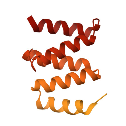
Chaperedoxin284Escherichia coli K-12Mutation(s): 0 
Gene Names: cnoXybbNb0492JW5067
UniProt
Find proteins for P77395 (Escherichia coli (strain K12))
Explore P77395 
Go to UniProtKB:  P77395
Entity Groups
Sequence Clusters30% Identity50% Identity70% Identity90% Identity95% Identity100% Identity
UniProt GroupP77395
Sequence Annotations
Expand
Reference Sequence
Find similar proteins by:|  3D Structure
Entity ID: 2
MoleculeChains  Sequence LengthOrganismDetailsImage
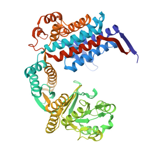
Chaperonin GroEL548Escherichia coli K-12Mutation(s): 0 
Gene Names: groELgroLmopAb4143JW4103
EC: 5.6.1.7
UniProt
Find proteins for P0A6F5 (Escherichia coli (strain K12))
Explore P0A6F5 
Go to UniProtKB:  P0A6F5
Entity Groups
Sequence Clusters30% Identity50% Identity70% Identity90% Identity95% Identity100% Identity
UniProt GroupP0A6F5
Sequence Annotations
Expand
Reference Sequence

Small Molecules

Ligands 3 Unique
IDChains Name / Formula / InChI Key2D Diagram3D Interactions
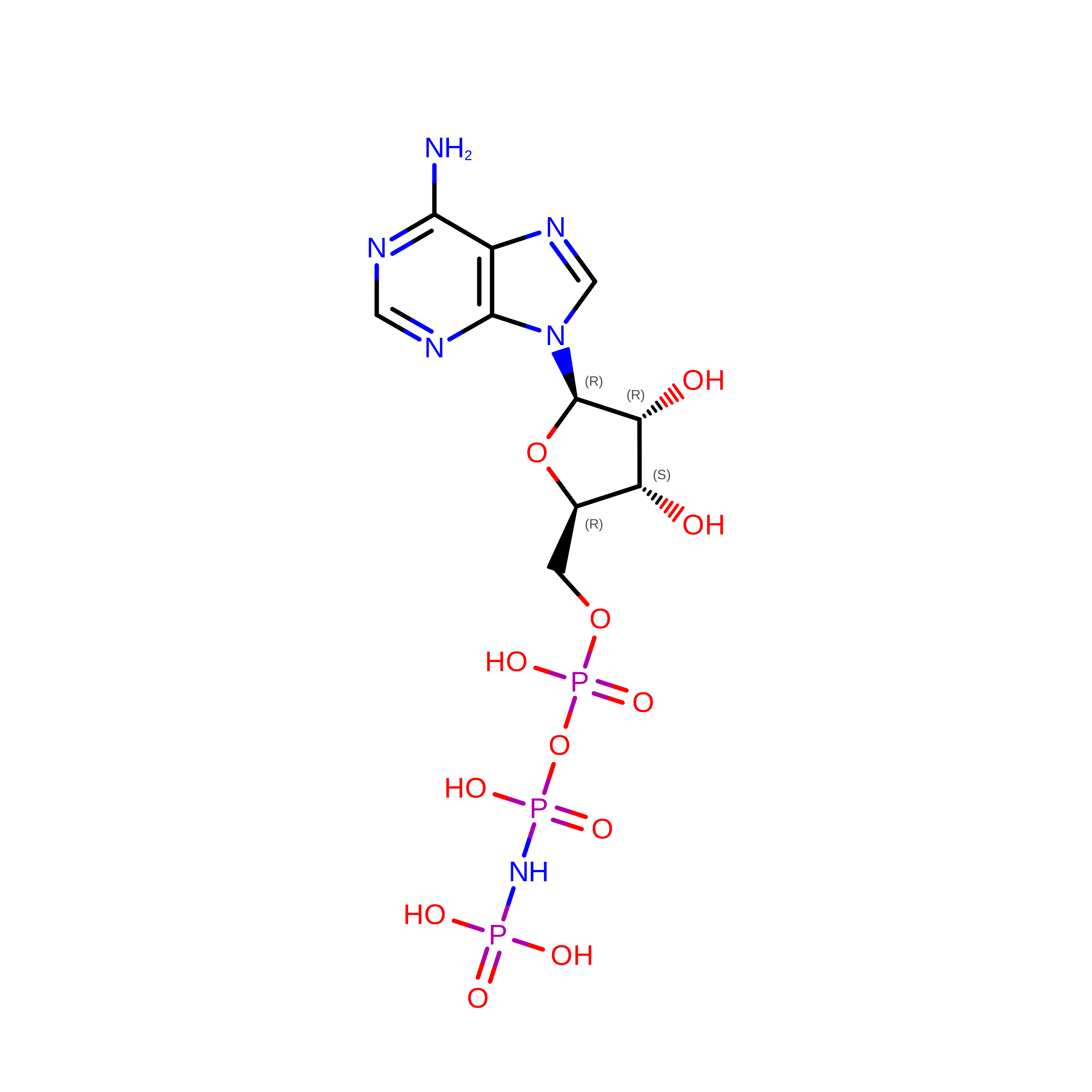
ANP
(Subject of Investigation/LOI)

Query on ANP


Download:Ideal Coordinates CCD File
CA [auth G]
CB [auth T]
EB [auth V]
HA [auth I]
HB [auth X]
CA [auth G],
CB [auth T],
EB [auth V],
HA [auth I],
HB [auth X],
KA [auth J],
LA [auth K],
LB [auth Z],
OB [auth b],
QA [auth L],
RA [auth N],
RB [auth c],
WA [auth P],
ZA [auth R]
PHOSPHOAMINOPHOSPHONIC ACID-ADENYLATE ESTER
C10 H17 N6 O12 P3
PVKSNHVPLWYQGJ-KQYNXXCUSA-N
K

Query on K


Download:Ideal Coordinates CCD File
AB [auth T]
DB [auth V]
EA [auth G]
GA [auth I]
GB [auth X]
AB [auth T],
DB [auth V],
EA [auth G],
GA [auth I],
GB [auth X],
IA [auth J],
KB [auth Z],
NA [auth K],
NB [auth b],
OA [auth L],
QB [auth c],
SA [auth N],
VA [auth P],
YA [auth R]
POTASSIUM ION
K
NPYPAHLBTDXSSS-UHFFFAOYSA-N
MG

Query on MG


Download:Ideal Coordinates CCD File
BB [auth T]
DA [auth G]
FA [auth I]
FB [auth V]
IB [auth X]
BB [auth T],
DA [auth G],
FA [auth I],
FB [auth V],
IB [auth X],
JA [auth J],
JB [auth Z],
MA [auth K],
MB [auth b],
PA [auth L],
PB [auth c],
TA [auth N],
UA [auth P],
XA [auth R]
MAGNESIUM ION
Mg
JLVVSXFLKOJNIY-UHFFFAOYSA-N

Experimental Data & Validation

Experimental Data

  • Method: ELECTRON MICROSCOPY
  • Resolution: 2.73 Å
  • Aggregation State: PARTICLE 
  • Reconstruction Method: SINGLE PARTICLE 
EM Software:
TaskSoftware PackageVersion
RECONSTRUCTIONcryoSPARC

Structure Validation

View Full Validation Report


Entry History 

& Funding Information

Deposition Data


Funding OrganizationLocationGrant Number
National Research Foundation (NRF, Korea)Korea, Republic Of--

Revision History  (Full details and data files)

  • Version 1.0: 2026-08-05
    Type: Initial release
  • Version 1.1: 2026-08-26
    Changes: Data collection, Database references