7QSQ

Permutated N-terminal lobe of the ribose binding protein from Thermotoga maritima


Experimental Data Snapshot

  • Method: X-RAY DIFFRACTION
  • Resolution: 1.79 Å
  • R-Value Free: 0.239 
  • R-Value Work: 0.191 
  • R-Value Observed: 0.193 

wwPDB Validation   3D Report Full Report


This is version 1.2 of the entry. See complete history


Literature

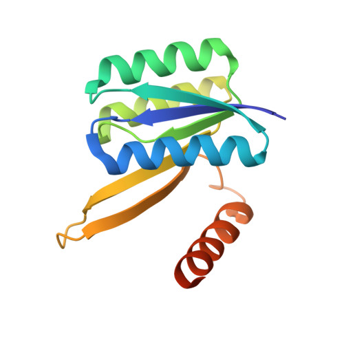
Structures of permuted halves of a modern ribose-binding protein.

Michel, F.Shanmugaratnam, S.Romero-Romero, S.Hocker, B.

(2023) Acta Crystallogr D Struct Biol 79: 40-49

  • DOI: https://doi.org/10.1107/S205979832201186X
  • Primary Citation of Related Structures:  
    7QSP, 7QSQ

  • PubMed Abstract: 

    Periplasmic binding proteins (PBPs) are a class of proteins that participate in the cellular transport of various ligands. They have been used as model systems to study mechanisms in protein evolution, such as duplication, recombination and domain swapping. It has been suggested that PBPs evolved from precursors half their size. Here, the crystal structures of two permuted halves of a modern ribose-binding protein (RBP) from Thermotoga maritima are reported. The overexpressed proteins are well folded and show a monomer-dimer equilibrium in solution. Their crystal structures show partially noncanonical PBP-like fold type I conformations with structural deviations from modern RBPs. One of the half variants forms a dimer via segment swapping, suggesting a high degree of malleability. The structural findings on these permuted halves support the evolutionary hypothesis that PBPs arose via a duplication event of a flavodoxin-like protein and further support a domain-swapping step that might have occurred during the evolution of the PBP-like fold, a process that is necessary to generate the characteristic motion of PBPs essential to perform their functions.


  • Organizational Affiliation

    Department of Biochemistry, University of Bayreuth, 95447 Bayreuth, Germany.


Macromolecules
Find similar proteins by:  (by identity cutoff)  |  3D Structure
Entity ID: 1
MoleculeChains Sequence LengthOrganismDetailsImage
Ribose ABC transporter, periplasmic ribose-binding proteinA,
B [auth C],
C [auth B],
D
138Thermotoga maritimaMutation(s): 0 
Gene Names: TM_0958
UniProt
Find proteins for Q9X053 (Thermotoga maritima (strain ATCC 43589 / DSM 3109 / JCM 10099 / NBRC 100826 / MSB8))
Explore Q9X053 
Go to UniProtKB:  Q9X053
Entity Groups  
Sequence Clusters30% Identity50% Identity70% Identity90% Identity95% Identity100% Identity
UniProt GroupQ9X053
Sequence Annotations
Expand
  • Reference Sequence
Small Molecules
Ligands 3 Unique
IDChains Name / Formula / InChI Key2D Diagram3D Interactions
PGE
Query on PGE

Download Ideal Coordinates CCD File 
M [auth C]TRIETHYLENE GLYCOL
C6 H14 O4
ZIBGPFATKBEMQZ-UHFFFAOYSA-N
SO4
Query on SO4

Download Ideal Coordinates CCD File 
E [auth A],
N [auth B]
SULFATE ION
O4 S
QAOWNCQODCNURD-UHFFFAOYSA-L
EDO
Query on EDO

Download Ideal Coordinates CCD File 
F [auth A]
G [auth A]
H [auth A]
I [auth A]
J [auth A]
F [auth A],
G [auth A],
H [auth A],
I [auth A],
J [auth A],
K [auth C],
L [auth C],
O [auth B],
P [auth B],
Q [auth D],
R [auth D],
S [auth D]
1,2-ETHANEDIOL
C2 H6 O2
LYCAIKOWRPUZTN-UHFFFAOYSA-N
Experimental Data & Validation

Experimental Data

  • Method: X-RAY DIFFRACTION
  • Resolution: 1.79 Å
  • R-Value Free: 0.239 
  • R-Value Work: 0.191 
  • R-Value Observed: 0.193 
  • Space Group: P 1 21 1
Unit Cell:
Length ( Å )Angle ( ˚ )
a = 55.37α = 90
b = 62.77β = 102.09
c = 76.26γ = 90
Software Package:
Software NamePurpose
PHENIXrefinement
XDSdata reduction
XDSdata scaling
PHASERphasing

Structure Validation

View Full Validation Report


Entry History & Funding Information

Deposition Data


Funding OrganizationLocationGrant Number
European Research Council (ERC)European Union647548

Revision History  (Full details and data files)

  • Version 1.0: 2023-01-11
    Type: Initial release
  • Version 1.1: 2023-01-25
    Changes: Database references
  • Version 1.2: 2024-01-31
    Changes: Data collection, Refinement description