7MWW

Structure of hepatitis C virus envelope full-length glycoprotein 2 (eE2) from J6 genotype


Experimental Data Snapshot

  • Method: X-RAY DIFFRACTION
  • Resolution: 2.71 Å
  • R-Value Free: 0.270 
  • R-Value Work: 0.218 
  • R-Value Observed: 0.221 

wwPDB Validation   3D Report Full Report


Ligand Structure Quality Assessment 


This is version 1.3 of the entry. See complete history


Literature

Structural insights into hepatitis C virus receptor binding and entry.

Kumar, A.Hossain, R.A.Yost, S.A.Bu, W.Wang, Y.Dearborn, A.D.Grakoui, A.Cohen, J.I.Marcotrigiano, J.

(2021) Nature 598: 521-525

  • DOI: https://doi.org/10.1038/s41586-021-03913-5
  • Primary Citation of Related Structures:  
    7MWS, 7MWW, 7MWX

  • PubMed Abstract: 

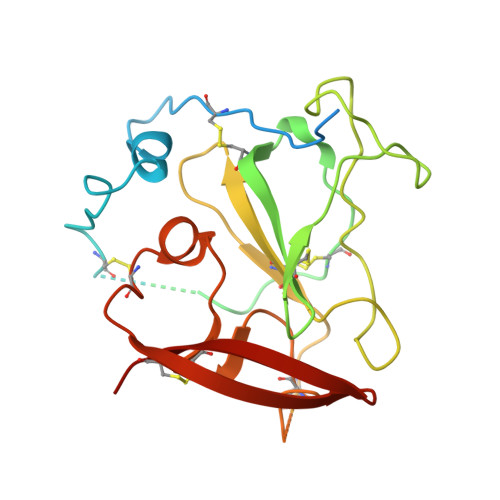
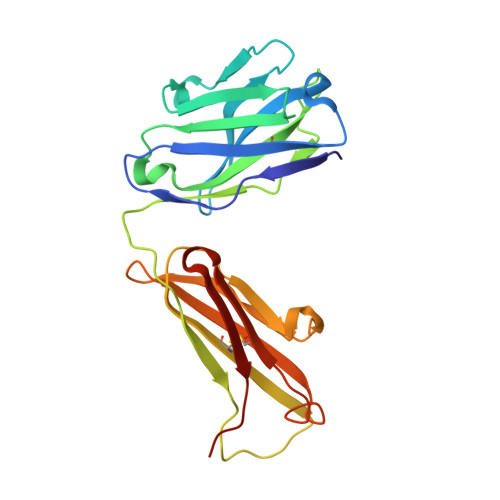
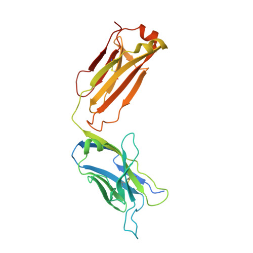
    Hepatitis C virus (HCV) infection is a causal agent of chronic liver disease, cirrhosis and hepatocellular carcinoma in humans, and afflicts more than 70 million people worldwide. The HCV envelope glycoproteins E1 and E2 are responsible for the binding of the virus to the host cell, but the exact entry process remains undetermined 1 . The majority of broadly neutralizing antibodies block interaction between HCV E2 and the large extracellular loop (LEL) of the cellular receptor CD81 (CD81-LEL) 2 . Here we show that low pH enhances the binding of CD81-LEL to E2, and we determine the crystal structure of E2 in complex with an antigen-binding fragment (2A12) and CD81-LEL (E2-2A12-CD81-LEL); E2 in complex with 2A12 (E2-2A12); and CD81-LEL alone. After binding CD81, residues 418-422 in E2 are displaced, which allows for the extension of an internal loop consisting of residues 520-539. Docking of the E2-CD81-LEL complex onto a membrane-embedded, full-length CD81 places the residues Tyr529 and Trp531 of E2 proximal to the membrane. Liposome flotation assays show that low pH and CD81-LEL increase the interaction of E2 with membranes, whereas structure-based mutants of Tyr529, Trp531 and Ile422 in the amino terminus of E2 abolish membrane binding. These data support a model in which acidification and receptor binding result in a conformational change in E2 in preparation for membrane fusion.


  • Organizational Affiliation

    Structural Virology Section, Laboratory of Infectious Diseases, National Institute of Allergy and Infectious Diseases, National Institutes of Health, Bethesda, MD, USA.


Macromolecules
Find similar proteins by:  (by identity cutoff)  |  3D Structure
Entity ID: 1
MoleculeChains Sequence LengthOrganismDetailsImage
Core protein precursor eE2A [auth E]272Hepacivirus hominisMutation(s): 0 
EC: 2.7.7.48 (PDB Primary Data), 3.4.21.98 (PDB Primary Data), 3.6.1.15 (PDB Primary Data), 3.6.4.13 (PDB Primary Data)
UniProt
Find proteins for A0A2I6PIY9 (Hepacivirus hominis)
Explore A0A2I6PIY9 
Go to UniProtKB:  A0A2I6PIY9
Entity Groups  
Sequence Clusters30% Identity50% Identity70% Identity90% Identity95% Identity100% Identity
UniProt GroupA0A2I6PIY9
Sequence Annotations
Expand
  • Reference Sequence
Find similar proteins by:  (by identity cutoff)  |  3D Structure
Entity ID: 2
MoleculeChains Sequence LengthOrganismDetailsImage
2A12 Fab Heavy chainB [auth H]221Mus musculusMutation(s): 0 
Entity Groups  
Sequence Clusters30% Identity50% Identity70% Identity90% Identity95% Identity100% Identity
Sequence Annotations
Expand
  • Reference Sequence
Find similar proteins by:  (by identity cutoff)  |  3D Structure
Entity ID: 3
MoleculeChains Sequence LengthOrganismDetailsImage
2A12 Fab light chainC [auth L]240Mus musculusMutation(s): 0 
Entity Groups  
Sequence Clusters30% Identity50% Identity70% Identity90% Identity95% Identity100% Identity
Sequence Annotations
Expand
  • Reference Sequence
Oligosaccharides

Help

Entity ID: 4
MoleculeChains Length2D Diagram Glycosylation3D Interactions
beta-D-mannopyranose-(1-4)-2-acetamido-2-deoxy-beta-D-glucopyranose-(1-4)-2-acetamido-2-deoxy-beta-D-glucopyranoseD [auth A]3N-Glycosylation
Glycosylation Resources
GlyTouCan:  G15407YE
GlyCosmos:  G15407YE
GlyGen:  G15407YE
Entity ID: 5
MoleculeChains Length2D Diagram Glycosylation3D Interactions
2-acetamido-2-deoxy-beta-D-glucopyranose-(1-4)-2-acetamido-2-deoxy-beta-D-glucopyranoseE [auth B]2N-Glycosylation
Glycosylation Resources
GlyTouCan:  G42666HT
GlyCosmos:  G42666HT
GlyGen:  G42666HT
Small Molecules
Experimental Data & Validation

Experimental Data

  • Method: X-RAY DIFFRACTION
  • Resolution: 2.71 Å
  • R-Value Free: 0.270 
  • R-Value Work: 0.218 
  • R-Value Observed: 0.221 
  • Space Group: C 2 2 21
Unit Cell:
Length ( Å )Angle ( ˚ )
a = 95.84α = 90
b = 155.561β = 90
c = 129.42γ = 90
Software Package:
Software NamePurpose
PHENIXrefinement
Cootmodel building
MOSFLMdata reduction
SCALAdata scaling
PHASERphasing

Structure Validation

View Full Validation Report


Ligand Structure Quality Assessment 


Entry History & Funding Information

Deposition Data


Funding OrganizationLocationGrant Number
National Institutes of Health/National Institute Of Allergy and Infectious Diseases (NIH/NIAID)United States--

Revision History  (Full details and data files)

  • Version 1.0: 2021-09-15
    Type: Initial release
  • Version 1.1: 2021-09-29
    Changes: Database references
  • Version 1.2: 2021-11-03
    Changes: Database references
  • Version 1.3: 2023-10-18
    Changes: Data collection, Refinement description