7MSK

ThuS glycosin S-glycosyltransferase


Experimental Data Snapshot

  • Method: X-RAY DIFFRACTION
  • Resolution: 2.06 Å
  • R-Value Free: 0.239 
  • R-Value Work: 0.201 
  • R-Value Observed: 0.202 

wwPDB Validation   3D Report Full Report


Ligand Structure Quality Assessment 


This is version 1.0 of the entry. See complete history


Literature

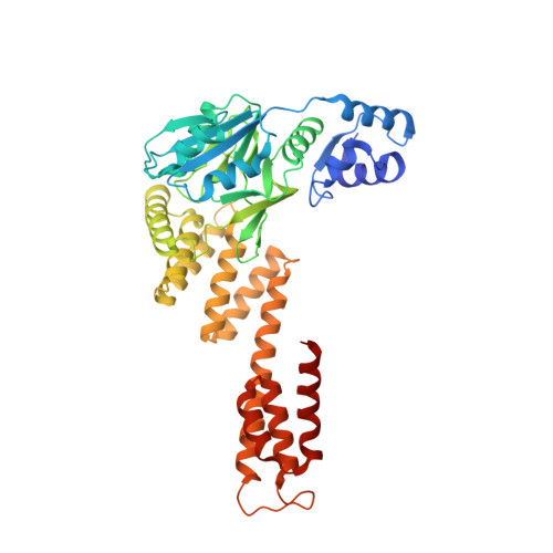
Structural and mechanistic investigations of protein S-glycosyltransferases.

Fujinami, D.Garcia de Gonzalo, C.V.Biswas, S.Hao, Y.Wang, H.Garg, N.Lukk, T.Nair, S.K.van der Donk, W.A.

(2021) Cell Chem Biol 28: 1740-1749.e6

  • DOI: https://doi.org/10.1016/j.chembiol.2021.06.009
  • Primary Citation of Related Structures:  
    7MSK, 7MSN, 7MSP

  • PubMed Abstract: 

    Attachment of sugars to nitrogen and oxygen in peptides is ubiquitous in biology, but glycosylation of sulfur atoms has only been recently described. Here, we characterize two S-glycosyltransferases SunS and ThuS that selectively glycosylate one of five Cys residues in their substrate peptides; substitution of this Cys with Ser results in a strong decrease in glycosylation activity. Crystal structures of SunS and ThuS in complex with UDP-glucose or a derivative reveal an unusual architecture in which a glycosyltransferase type A (GTA) fold is decorated with additional domains to support homodimerization. Dimer formation creates an extended cavity for the substrate peptide, drawing functional analogy with O-glycosyltransferases involved in cell wall biosynthesis. This extended cavity contains a sharp bend that may explain the site selectivity of the glycosylation because the target Cys is in a Gly-rich stretch that can accommodate the bend. These studies establish a molecular framework for understanding the unusual S-glycosyltransferases.


  • Organizational Affiliation

    Howard Hughes Medical Institute and Roger Adams Laboratory, Department of Chemistry, University of Illinois at Urbana-Champaign, 600 South Mathews Avenue, Urbana, IL 61801, USA.


Macromolecules
Find similar proteins by:  (by identity cutoff)  |  3D Structure
Entity ID: 1
MoleculeChains Sequence LengthOrganismDetailsImage
Glyco_trans_2-like domain-containing protein
A, B
430Bacillus thuringiensis serovar andalousiensis BGSC 4AW1Mutation(s): 0 
Gene Names: bthur0009_56280
UniProt
Find proteins for C3GCL5 (Bacillus thuringiensis serovar andalousiensis BGSC 4AW1)
Explore C3GCL5 
Go to UniProtKB:  C3GCL5
Entity Groups  
Sequence Clusters30% Identity50% Identity70% Identity90% Identity95% Identity100% Identity
UniProt GroupC3GCL5
Sequence Annotations
Expand
  • Reference Sequence
Experimental Data & Validation

Experimental Data

  • Method: X-RAY DIFFRACTION
  • Resolution: 2.06 Å
  • R-Value Free: 0.239 
  • R-Value Work: 0.201 
  • R-Value Observed: 0.202 
  • Space Group: P 1 21 1
Unit Cell:
Length ( Å )Angle ( ˚ )
a = 49.833α = 90
b = 112.654β = 90.11
c = 90.31γ = 90
Software Package:
Software NamePurpose
REFMACrefinement
PDB_EXTRACTdata extraction
autoPROCdata reduction
Aimlessdata scaling
PHASERphasing

Structure Validation

View Full Validation Report


Ligand Structure Quality Assessment 


Entry History & Funding Information

Deposition Data


Funding OrganizationLocationGrant Number
National Institutes of Health/National Institute of General Medical Sciences (NIH/NIGMS)United StatesGM079038

Revision History  (Full details and data files)

  • Version 1.0: 2022-04-13
    Type: Initial release