7EHN

Human MTHFD2 in complex with compound 21 and 9


Experimental Data Snapshot

  • Method: X-RAY DIFFRACTION
  • Resolution: 2.25 Å
  • R-Value Free: 0.240 
  • R-Value Work: 0.201 
  • R-Value Observed: 0.203 

wwPDB Validation   3D Report Full Report


Ligand Structure Quality Assessment 


This is version 1.2 of the entry. See complete history


Literature

Xanthine Derivatives Reveal an Allosteric Binding Site in Methylenetetrahydrofolate Dehydrogenase 2 (MTHFD2).

Lee, L.C.Peng, Y.H.Chang, H.H.Hsu, T.Lu, C.T.Huang, C.H.Hsueh, C.C.Kung, F.C.Kuo, C.C.Jiaang, W.T.Wu, S.Y.

(2021) J Med Chem 64: 11288-11301

  • DOI: https://doi.org/10.1021/acs.jmedchem.1c00663
  • Primary Citation of Related Structures:  
    7EHJ, 7EHM, 7EHN, 7EHV

  • PubMed Abstract: 

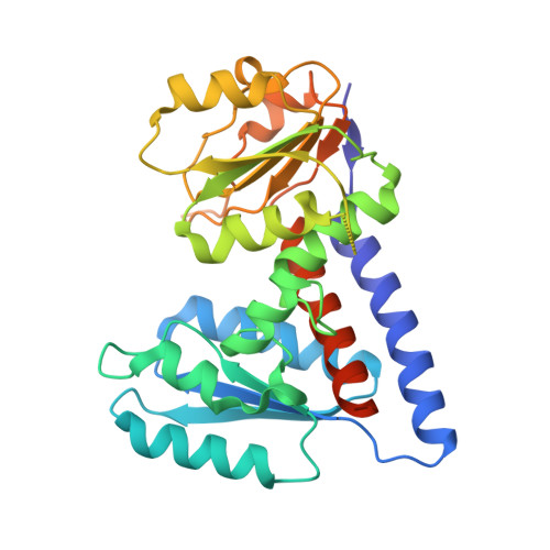
    Methylenetetrahydrofolate dehydrogenase 2 (MTHFD2) plays an important role in one-carbon metabolism. The MTHFD2 gene is upregulated in various cancers but very low or undetectable in normal proliferating cells, and therefore a potential target for cancer treatment. In this study, we present the structure of MTHFD2 in complex with xanthine derivative 15 , which allosterically binds to MTHFD2 and coexists with the substrate analogue. A kinetic study demonstrated the uncompetitive inhibition of MTHFD2 by 15 . Allosteric inhibitors often provide good selectivity and, indeed, xanthine derivatives are highly selective for MTHFD2. Moreover, several conformational changes were observed upon the binding of 15 , which impeded the binding of the cofactor and phosphate to MTHFD2. To the best of our knowledge, this is the first study to identify allosteric inhibitors targeting the MTHFD family and our results would provide insights on the inhibition mechanism of MTHFD proteins and the development of novel inhibitors.


  • Organizational Affiliation

    Institute of Biotechnology and Pharmaceutical Research, National Health Research Institutes, 35, Keyan Road, Zhunan Town, Miaoli County 350 Taiwan, ROC.


Macromolecules
Find similar proteins by:  (by identity cutoff)  |  3D Structure
Entity ID: 1
MoleculeChains Sequence LengthOrganismDetailsImage
Bifunctional methylenetetrahydrofolate dehydrogenase/cyclohydrolase, mitochondrial
A, B
315Homo sapiensMutation(s): 0 
Gene Names: MTHFD2NMDMC
EC: 1.5.1.15 (PDB Primary Data), 3.5.4.9 (PDB Primary Data)
UniProt & NIH Common Fund Data Resources
Find proteins for P13995 (Homo sapiens)
Explore P13995 
Go to UniProtKB:  P13995
PHAROS:  P13995
GTEx:  ENSG00000065911 
Entity Groups  
Sequence Clusters30% Identity50% Identity70% Identity90% Identity95% Identity100% Identity
UniProt GroupP13995
Sequence Annotations
Expand
  • Reference Sequence
Small Molecules
Ligands 2 Unique
IDChains Name / Formula / InChI Key2D Diagram3D Interactions
J4F (Subject of Investigation/LOI)
Query on J4F

Download Ideal Coordinates CCD File 
C [auth A],
E [auth B]
3-[4-[[1-[(4-chloranyl-1H-indol-2-yl)methyl]-3,7-dimethyl-2,6-bis(oxidanylidene)purin-8-yl]amino]-6-methyl-pyrimidin-2-yl]propanoic acid
C24 H23 Cl N8 O4
ILEKCBRCJDWEIR-UHFFFAOYSA-N
J49
Query on J49

Download Ideal Coordinates CCD File 
D [auth A],
F [auth B]
(2S)-2-[[4-[(4-azanyl-6-oxidanyl-pyrimidin-5-yl)carbamoylamino]phenyl]carbonylamino]pentanedioic acid
C17 H18 N6 O7
OUMOQELZARCDHX-JTQLQIEISA-N
Experimental Data & Validation

Experimental Data

  • Method: X-RAY DIFFRACTION
  • Resolution: 2.25 Å
  • R-Value Free: 0.240 
  • R-Value Work: 0.201 
  • R-Value Observed: 0.203 
  • Space Group: P 65
Unit Cell:
Length ( Å )Angle ( ˚ )
a = 113.169α = 90
b = 113.169β = 90
c = 113.67γ = 120
Software Package:
Software NamePurpose
PHENIXrefinement
HKL-2000data reduction
SCALEPACKdata scaling
MOLREPphasing
PDB_EXTRACTdata extraction

Structure Validation

View Full Validation Report


Ligand Structure Quality Assessment 


Entry History 

Deposition Data

Revision History  (Full details and data files)

  • Version 1.0: 2021-08-11
    Type: Initial release
  • Version 1.1: 2022-02-16
    Changes: Database references
  • Version 1.2: 2023-11-29
    Changes: Data collection, Refinement description