7BR3

Crystal structure of the protein 1


Experimental Data Snapshot

  • Method: X-RAY DIFFRACTION
  • Resolution: 2.79 Å
  • R-Value Free: 0.284 
  • R-Value Work: 0.243 
  • R-Value Observed: 0.247 

wwPDB Validation   3D Report Full Report


Ligand Structure Quality Assessment 


This is version 1.2 of the entry. See complete history


Literature

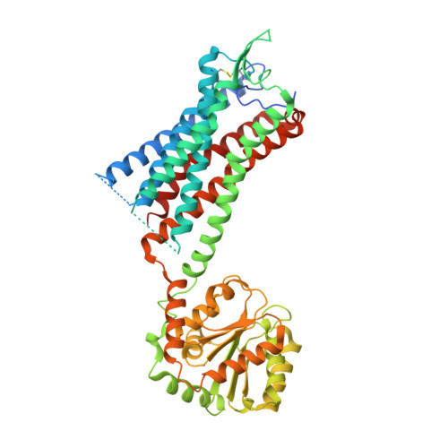
Structure of the human gonadotropin-releasing hormone receptor GnRH1R reveals an unusual ligand binding mode.

Yan, W.Cheng, L.Wang, W.Wu, C.Yang, X.Du, X.Ma, L.Qi, S.Wei, Y.Lu, Z.Yang, S.Shao, Z.

(2020) Nat Commun 11: 5287-5287

  • DOI: https://doi.org/10.1038/s41467-020-19109-w
  • Primary Citation of Related Structures:  
    7BR3

  • PubMed Abstract: 

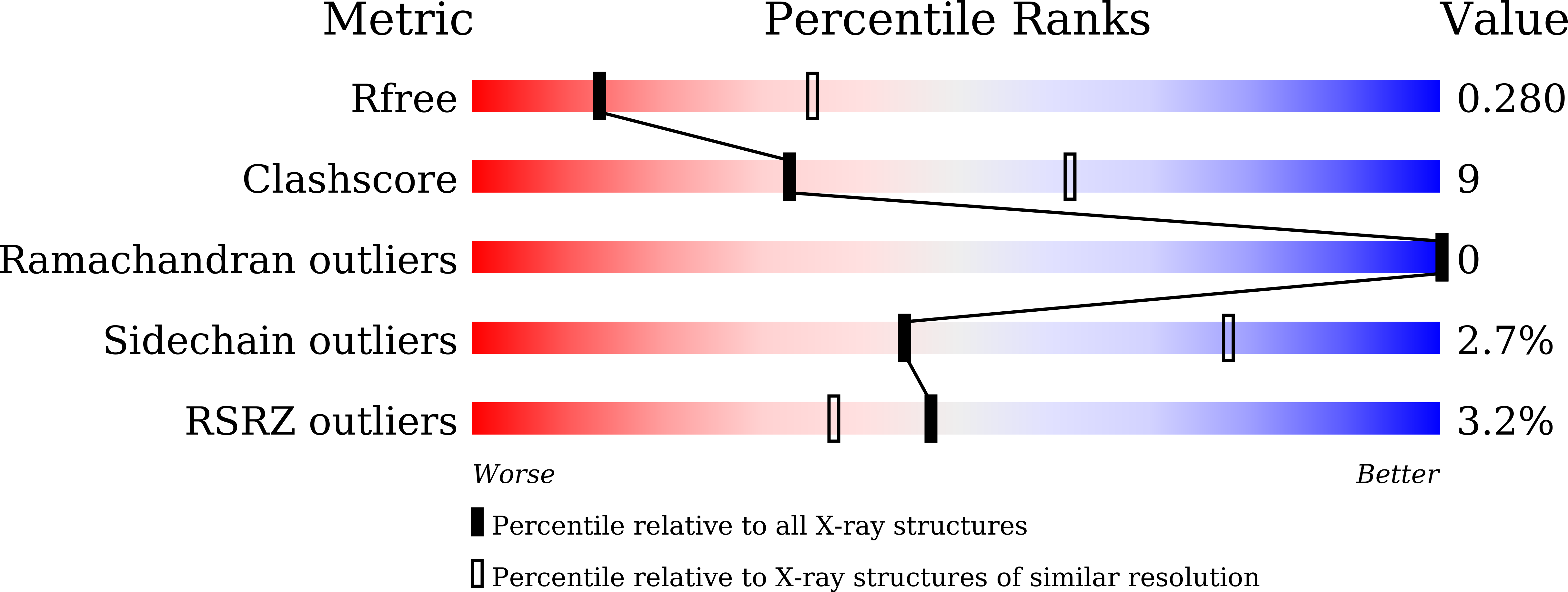
    Gonadotrophin-releasing hormone (GnRH), also known as luteinizing hormone-releasing hormone, is the main regulator of the reproductive system, acting on gonadotropic cells by binding to the GnRH1 receptor (GnRH1R). The GnRH-GnRH1R system is a promising therapeutic target for maintaining reproductive function; to date, a number of ligands targeting GnRH1R for disease treatment are available on the market. Here, we report the crystal structure of GnRH1R bound to the small-molecule drug elagolix at 2.8 Å resolution. The structure reveals an interesting N-terminus that could co-occupy the enlarged orthosteric binding site together with elagolix. The unusual ligand binding mode was further investigated by structural analyses, functional assays and molecular docking studies. On the other hand, because of the unique characteristic of lacking a cytoplasmic C-terminal helix, GnRH1R exhibits different microswitch structural features from other class A GPCRs. In summary, this study provides insight into the ligand binding mode of GnRH1R and offers an atomic framework for rational drug design.


  • Organizational Affiliation

    Division of Nephrology and Kidney Research Institute, State Key Laboratory of Biotherapy and Cancer Center, West China Hospital, Sichuan University, Chengdu, Sichuan, 610041, China.


Macromolecules
Find similar proteins by:  (by identity cutoff)  |  3D Structure
Entity ID: 1
MoleculeChains Sequence LengthOrganismDetailsImage
Gonadotropin-releasing hormone receptor,GlgA glycogen synthase,Gonadotropin-releasing hormone receptor528Homo sapiensPyrococcus abyssi GE5Mutation(s): 1 
Gene Names: GNRHRGRHRPAB2292
Membrane Entity: Yes 
UniProt & NIH Common Fund Data Resources
Find proteins for Q9V2J8 (Pyrococcus abyssi (strain GE5 / Orsay))
Explore Q9V2J8 
Go to UniProtKB:  Q9V2J8
Find proteins for P30968 (Homo sapiens)
Explore P30968 
Go to UniProtKB:  P30968
PHAROS:  P30968
GTEx:  ENSG00000109163 
Entity Groups  
Sequence Clusters30% Identity50% Identity70% Identity90% Identity95% Identity100% Identity
UniProt GroupsQ9V2J8P30968
Sequence Annotations
Expand
  • Reference Sequence
Small Molecules
Ligands 5 Unique
IDChains Name / Formula / InChI Key2D Diagram3D Interactions
F5O (Subject of Investigation/LOI)
Query on F5O

Download Ideal Coordinates CCD File 
L [auth A]4-[[(1R)-2-[5-(2-fluoranyl-3-methoxy-phenyl)-3-[[2-fluoranyl-6-(trifluoromethyl)phenyl]methyl]-4-methyl-2,6-bis(oxidanylidene)pyrimidin-1-yl]-1-phenyl-ethyl]amino]butanoic acid
C32 H30 F5 N3 O5
HEAUOKZIVMZVQL-VWLOTQADSA-N
OLC (Subject of Investigation/LOI)
Query on OLC

Download Ideal Coordinates CCD File 
C [auth A],
D [auth A]
(2R)-2,3-dihydroxypropyl (9Z)-octadec-9-enoate
C21 H40 O4
RZRNAYUHWVFMIP-GDCKJWNLSA-N
1QW
Query on 1QW

Download Ideal Coordinates CCD File 
B [auth A](2R)-2,3-dihydroxypropyl dodecanoate
C15 H30 O4
ARIWANIATODDMH-CQSZACIVSA-N
PEG
Query on PEG

Download Ideal Coordinates CCD File 
E [auth A]
F [auth A]
G [auth A]
H [auth A]
I [auth A]
E [auth A],
F [auth A],
G [auth A],
H [auth A],
I [auth A],
J [auth A]
DI(HYDROXYETHYL)ETHER
C4 H10 O3
MTHSVFCYNBDYFN-UHFFFAOYSA-N
FMT
Query on FMT

Download Ideal Coordinates CCD File 
K [auth A]FORMIC ACID
C H2 O2
BDAGIHXWWSANSR-UHFFFAOYSA-N
Experimental Data & Validation

Experimental Data

  • Method: X-RAY DIFFRACTION
  • Resolution: 2.79 Å
  • R-Value Free: 0.284 
  • R-Value Work: 0.243 
  • R-Value Observed: 0.247 
  • Space Group: P 31 2 1
Unit Cell:
Length ( Å )Angle ( ˚ )
a = 73.964α = 90
b = 73.964β = 90
c = 231.156γ = 120
Software Package:
Software NamePurpose
PHENIXrefinement
SCALAdata scaling
PDB_EXTRACTdata extraction
HKL-3000data reduction
PHENIXphasing

Structure Validation

View Full Validation Report


Ligand Structure Quality Assessment 


Entry History 

Deposition Data

Revision History  (Full details and data files)

  • Version 1.0: 2020-10-07
    Type: Initial release
  • Version 1.1: 2020-11-04
    Changes: Database references
  • Version 1.2: 2023-11-29
    Changes: Data collection, Database references, Refinement description