7B26

CirpA1 in complex with pseudo-monomeric Properdin lacking TSR2-3


Experimental Data Snapshot

  • Method: X-RAY DIFFRACTION
  • Resolution: 3.40 Å
  • R-Value Free: 0.274 
  • R-Value Work: 0.229 
  • R-Value Observed: 0.232 

wwPDB Validation   3D Report Full Report


This is version 1.2 of the entry. See complete history


Literature

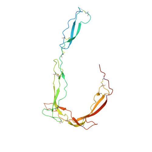
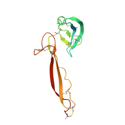
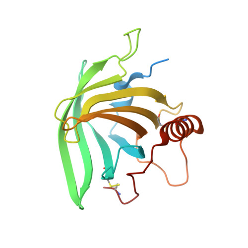
Structure and function of a family of tick-derived complement inhibitors targeting properdin.

Braunger, K.Ahn, J.Jore, M.M.Johnson, S.Tang, T.T.L.Pedersen, D.V.Andersen, G.R.Lea, S.M.

(2022) Nat Commun 13: 317-317

  • DOI: https://doi.org/10.1038/s41467-021-27920-2
  • Primary Citation of Related Structures:  
    7B26, 7B28, 7B29, 7B2A, 7B2D

  • PubMed Abstract: 

    Activation of the serum-resident complement system begins a cascade that leads to activation of membrane-resident complement receptors on immune cells, thus coordinating serum and cellular immune responses. Whilst many molecules act to control inappropriate activation, Properdin is the only known positive regulator of the human complement system. By stabilising the alternative pathway C3 convertase it promotes complement self-amplification and persistent activation boosting the magnitude of the serum complement response by all triggers. In this work, we identify a family of tick-derived alternative pathway complement inhibitors, hereafter termed CirpA. Functional and structural characterisation reveals that members of the CirpA family directly bind to properdin, inhibiting its ability to promote complement activation, and leading to potent inhibition of the complement response in a species specific manner. We provide a full functional and structural characterisation of a properdin inhibitor, opening avenues for future therapeutic approaches.


  • Organizational Affiliation

    Sir William Dunn School of Pathology, University of Oxford, OX1 3RE, Oxford, UK.


Macromolecules
Find similar proteins by:  (by identity cutoff)  |  3D Structure
Entity ID: 1
MoleculeChains Sequence LengthOrganismDetailsImage
ProperdinA [auth B]247Homo sapiensMutation(s): 0 
Gene Names: CFPPFC
UniProt & NIH Common Fund Data Resources
Find proteins for P27918 (Homo sapiens)
Explore P27918 
Go to UniProtKB:  P27918
PHAROS:  P27918
GTEx:  ENSG00000126759 
Entity Groups  
Sequence Clusters30% Identity50% Identity70% Identity90% Identity95% Identity100% Identity
UniProt GroupP27918
Sequence Annotations
Expand
  • Reference Sequence
Find similar proteins by:  (by identity cutoff)  |  3D Structure
Entity ID: 2
MoleculeChains Sequence LengthOrganismDetailsImage
ProperdinB [auth A]134Homo sapiensMutation(s): 1 
Gene Names: CFPPFC
UniProt & NIH Common Fund Data Resources
Find proteins for P27918 (Homo sapiens)
Explore P27918 
Go to UniProtKB:  P27918
PHAROS:  P27918
GTEx:  ENSG00000126759 
Entity Groups  
Sequence Clusters30% Identity50% Identity70% Identity90% Identity95% Identity100% Identity
UniProt GroupP27918
Sequence Annotations
Expand
  • Reference Sequence
Find similar proteins by:  (by identity cutoff)  |  3D Structure
Entity ID: 3
MoleculeChains Sequence LengthOrganismDetailsImage
CirpA1180Rhipicephalus pulchellusMutation(s): 0 
Entity Groups  
Sequence Clusters30% Identity50% Identity70% Identity90% Identity95% Identity100% Identity
Sequence Annotations
Expand
  • Reference Sequence
Oligosaccharides

Help

Entity ID: 4
MoleculeChains Length2D Diagram Glycosylation3D Interactions
beta-D-glucopyranose-(1-3)-alpha-L-fucopyranose
D
2O-Glycosylation
Glycosylation Resources
GlyTouCan:  G36855WW
GlyCosmos:  G36855WW
GlyGen:  G36855WW
Small Molecules
Ligands 2 Unique
IDChains Name / Formula / InChI Key2D Diagram3D Interactions
BMA
Query on BMA

Download Ideal Coordinates CCD File 
J [auth B]beta-D-mannopyranose
C6 H12 O6
WQZGKKKJIJFFOK-RWOPYEJCSA-N
MAN
Query on MAN

Download Ideal Coordinates CCD File 
E [auth B]
F [auth B]
G [auth B]
H [auth B]
I [auth B]
E [auth B],
F [auth B],
G [auth B],
H [auth B],
I [auth B],
K [auth B],
L [auth A],
M [auth A]
alpha-D-mannopyranose
C6 H12 O6
WQZGKKKJIJFFOK-PQMKYFCFSA-N
Experimental Data & Validation

Experimental Data

  • Method: X-RAY DIFFRACTION
  • Resolution: 3.40 Å
  • R-Value Free: 0.274 
  • R-Value Work: 0.229 
  • R-Value Observed: 0.232 
  • Space Group: P 21 21 2
Unit Cell:
Length ( Å )Angle ( ˚ )
a = 166.31α = 90
b = 54.512β = 90
c = 70.581γ = 90
Software Package:
Software NamePurpose
XDSdata reduction
Aimlessdata scaling
MOLREPphasing
Cootmodel building
PHENIXrefinement

Structure Validation

View Full Validation Report


Entry History & Funding Information

Deposition Data


Funding OrganizationLocationGrant Number
Wellcome TrustUnited Kingdom100298
Wellcome TrustUnited Kingdom209194
Wellcome TrustUnited Kingdom219477
Medical Research Council (MRC, United Kingdom)United KingdomS021264
European Molecular Biology Organization (EMBO)United KingdomALTF 554-2019

Revision History  (Full details and data files)

  • Version 1.0: 2021-12-08
    Type: Initial release
  • Version 1.1: 2022-02-02
    Changes: Database references
  • Version 1.2: 2024-01-31
    Changes: Data collection, Refinement description