7AXZ

Ku70/80 complex apo form


Experimental Data Snapshot

  • Method: ELECTRON MICROSCOPY
  • Resolution: 3.20 Å
  • Aggregation State: PARTICLE 
  • Reconstruction Method: SINGLE PARTICLE 

wwPDB Validation   3D Report Full Report


This is version 1.1 of the entry. See complete history


Literature

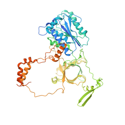
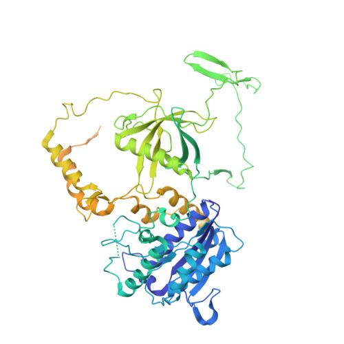
SAP domain forms a flexible part of DNA aperture in Ku70/80.

Hnizda, A.Tesina, P.Nguyen, T.B.Kukacka, Z.Kater, L.Chaplin, A.K.Beckmann, R.Ascher, D.B.Novak, P.Blundell, T.L.

(2021) FEBS J 288: 4382-4393

  • DOI: https://doi.org/10.1111/febs.15732
  • Primary Citation of Related Structures:  
    7AXZ

  • PubMed Abstract: 

    Nonhomologous end joining (NHEJ) is a DNA repair mechanism that religates double-strand DNA breaks to maintain genomic integrity during the entire cell cycle. The Ku70/80 complex recognizes DNA breaks and serves as an essential hub for recruitment of NHEJ components. Here, we describe intramolecular interactions of the Ku70 C-terminal domain, known as the SAP domain. Using single-particle cryo-electron microscopy, mass spectrometric analysis of intermolecular cross-linking and molecular modelling simulations, we captured variable positions of the SAP domain depending on DNA binding. The first position was localized at the DNA aperture in the Ku70/80 apo form but was not observed in the DNA-bound state. The second position, which was observed in both apo and DNA-bound states, was found below the DNA aperture, close to the helical arm of Ku70. The localization of the SAP domain in the DNA aperture suggests a function as a flexible entry gate for broken DNA. DATABASES: EM maps have been deposited in EMDB (EMD-11933). Coordinates have been deposited in Protein Data Bank (PDB 7AXZ). Other data are available from corresponding authors upon a request.


  • Organizational Affiliation

    Department of Biochemistry, University of Cambridge, Cambridge, UK.


Macromolecules
Find similar proteins by:  (by identity cutoff)  |  3D Structure
Entity ID: 1
MoleculeChains Sequence LengthOrganismDetailsImage
X-ray repair cross-complementing protein 6609Homo sapiensMutation(s): 0 
Gene Names: XRCC6G22P1
EC: 3.6.4 (PDB Primary Data), 4.2.99 (PDB Primary Data)
UniProt & NIH Common Fund Data Resources
Find proteins for P12956 (Homo sapiens)
Explore P12956 
Go to UniProtKB:  P12956
PHAROS:  P12956
GTEx:  ENSG00000196419 
Entity Groups  
Sequence Clusters30% Identity50% Identity70% Identity90% Identity95% Identity100% Identity
UniProt GroupP12956
Sequence Annotations
Expand
  • Reference Sequence
Find similar proteins by:  (by identity cutoff)  |  3D Structure
Entity ID: 2
MoleculeChains Sequence LengthOrganismDetailsImage
X-ray repair cross-complementing protein 5732Homo sapiensMutation(s): 0 
Gene Names: XRCC5G22P2
EC: 3.6.4
UniProt & NIH Common Fund Data Resources
Find proteins for P13010 (Homo sapiens)
Explore P13010 
Go to UniProtKB:  P13010
PHAROS:  P13010
GTEx:  ENSG00000079246 
Entity Groups  
Sequence Clusters30% Identity50% Identity70% Identity90% Identity95% Identity100% Identity
UniProt GroupP13010
Sequence Annotations
Expand
  • Reference Sequence
Experimental Data & Validation

Experimental Data

  • Method: ELECTRON MICROSCOPY
  • Resolution: 3.20 Å
  • Aggregation State: PARTICLE 
  • Reconstruction Method: SINGLE PARTICLE 

Structure Validation

View Full Validation Report


Entry History & Funding Information

Deposition Data


Funding OrganizationLocationGrant Number
Wellcome TrustUnited Kingdom200814/Z/16/Z; 2016

Revision History  (Full details and data files)

  • Version 1.0: 2021-02-10
    Type: Initial release
  • Version 1.1: 2021-07-28
    Changes: Database references