


Mechanistic insights into carbapenem hydrolysis by OXA-48 and the OXA10-derived hybrids OXA-10 loop24 and loop48
Tassone, G., Di Pisa, F., Benvenuti, M., De Luca, F., Pozzi, C., Mangani, S., Docquier, J.D.To be published.
Experimental Data Snapshot
Entity ID: 1 | |||||
|---|---|---|---|---|---|
| Molecule | Chains | Sequence Length | Organism | Details | Image |
| Beta-lactamase | 243 | Escherichia coli | Mutation(s): 0 Gene Names: blaoxa-10, blaOXA-10, oxa-10, BK373_28375, CQP61_30695, E4K55_27185, FORC82_p097, GII67_09965 EC: 3.5.2.6 | 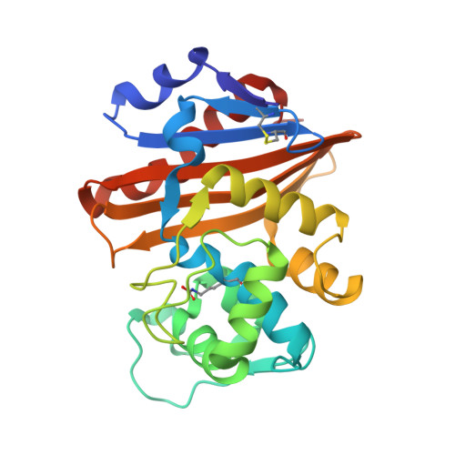 | |
UniProt | |||||
Find proteins for Q7BNC2 (Escherichia coli) Explore Q7BNC2 Go to UniProtKB: Q7BNC2 | |||||
Entity Groups | |||||
| Sequence Clusters | 30% Identity50% Identity70% Identity90% Identity95% Identity100% Identity | ||||
| UniProt Group | Q7BNC2 | ||||
Sequence AnnotationsExpand | |||||
| |||||
| Ligands 2 Unique | |||||
|---|---|---|---|---|---|
| ID | Chains | Name / Formula / InChI Key | 2D Diagram | 3D Interactions | |
| DWZ (Subject of Investigation/LOI) Query on DWZ | G [auth A], K [auth B], P [auth C], S [auth D] | (2S,3R,4S)-4-{[(3S,5S)-5-(dimethylcarbamoyl)pyrrolidin-3-yl]sulfanyl}-2-[(2S,3R)-3-hydroxy-1-oxobutan-2-yl]-3-methyl-3,4-dihydro-2H-pyrrole-5-carboxylic acid C17 H27 N3 O5 S UUIYVKJXUXGPKB-VGWSNGFZSA-N |  | ||
| EDO Query on EDO | E [auth A] F [auth A] H [auth A] I [auth A] J [auth B] | 1,2-ETHANEDIOL C2 H6 O2 LYCAIKOWRPUZTN-UHFFFAOYSA-N |  | ||
| Modified Residues 1 Unique | |||||
|---|---|---|---|---|---|
| ID | Chains | Type | Formula | 2D Diagram | Parent |
| KCX Query on KCX | A, B, C, D | L-PEPTIDE LINKING | C7 H14 N2 O4 |  | LYS |
| Length ( Å ) | Angle ( ˚ ) |
|---|---|
| a = 80.64 | α = 90 |
| b = 80.64 | β = 90 |
| c = 152.29 | γ = 120 |
| Software Name | Purpose |
|---|---|
| REFMAC | refinement |
| PDB_EXTRACT | data extraction |
| iMOSFLM | data reduction |
| SCALA | data scaling |
| MOLREP | phasing |
RCSB PDB (citation) is hosted by
RCSB PDB is a member of the

