6WEV

Crystal structures of human E-NPP 1: bound to N-{[1-(6,7-dimethoxy-5,8-dihydroquinazolin-4-yl)piperidin-4-yl]methyl}sulfuric diamide


Experimental Data Snapshot

  • Method: X-RAY DIFFRACTION
  • Resolution: 2.90 Å
  • R-Value Free: 0.253 
  • R-Value Work: 0.211 

wwPDB Validation   3D Report Full Report


Ligand Structure Quality Assessment 


This is version 1.2 of the entry. See complete history


Literature

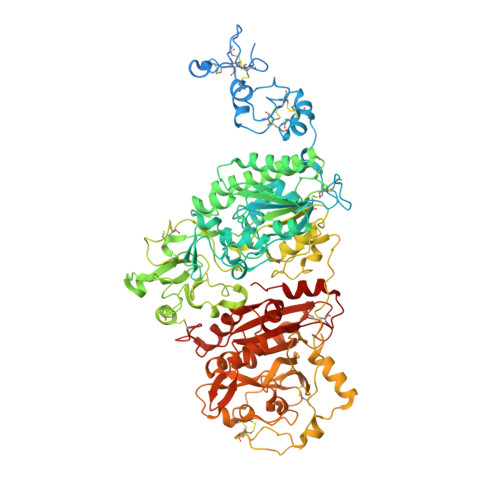
Crystal structures of human ENPP1 in apo and bound forms.

Dennis, M.L.Newman, J.Dolezal, O.Hattarki, M.Surjadi, R.N.Nuttall, S.D.Pham, T.Nebl, T.Camerino, M.Khoo, P.S.Monahan, B.J.Peat, T.S.

(2020) Acta Crystallogr D Struct Biol 76: 889-898

  • DOI: https://doi.org/10.1107/S2059798320010505
  • Primary Citation of Related Structures:  
    6WET, 6WEU, 6WEV, 6WEW, 6WFJ

  • PubMed Abstract: 

    Cancer is one of the leading causes of mortality in humans, and recent work has focused on the area of immuno-oncology, in which the immune system is used to specifically target cancerous cells. Ectonucleotide pyrophosphatase/phosphodiesterase 1 (ENPP1) is an emerging therapeutic target in human cancers owing to its role in degrading cyclic GMP-AMP (cGAMP), an agonist of the stimulator of interferon genes (STING). The available structures of ENPP1 are of the mouse enzyme, and no structures are available with anything other than native nucleotides. Here, the first X-ray crystal structures of the human ENPP1 enzyme in an apo form, with bound nucleotides and with two known inhibitors are presented. The availability of these structures and a robust crystallization system will allow the development of structure-based drug-design campaigns against this attractive cancer therapeutic target.


  • Organizational Affiliation

    Biomedical Manufacturing Program, CSIRO, 343 Royal Parade, Parkville, VIC 3052, Australia.


Macromolecules
Find similar proteins by:  (by identity cutoff)  |  3D Structure
Entity ID: 1
MoleculeChains Sequence LengthOrganismDetailsImage
Ectonucleotide pyrophosphatase/phosphodiesterase family member 1A [auth AbA],
B [auth BbB]
925Homo sapiensMutation(s): 0 
Gene Names: ENPP1M6S1NPPSPC1PDNP1
EC: 3.1.4.1 (PDB Primary Data), 3.6.1.9 (PDB Primary Data)
UniProt & NIH Common Fund Data Resources
Find proteins for P22413 (Homo sapiens)
Explore P22413 
Go to UniProtKB:  P22413
PHAROS:  P22413
GTEx:  ENSG00000197594 
Entity Groups  
Sequence Clusters30% Identity50% Identity70% Identity90% Identity95% Identity100% Identity
UniProt GroupP22413
Sequence Annotations
Expand
  • Reference Sequence
Oligosaccharides

Help

Entity ID: 2
MoleculeChains Length2D Diagram Glycosylation3D Interactions
alpha-D-mannopyranose-(1-3)-alpha-D-mannopyranose-(1-4)-2-acetamido-2-deoxy-beta-D-glucopyranose-(1-4)-2-acetamido-2-deoxy-beta-D-glucopyranoseC [auth AcA]4N-Glycosylation
Glycosylation Resources
GlyTouCan:  G48068RF
GlyCosmos:  G48068RF
GlyGen:  G48068RF
Entity ID: 3
MoleculeChains Length2D Diagram Glycosylation3D Interactions
beta-D-mannopyranose-(1-4)-2-acetamido-2-deoxy-beta-D-glucopyranose-(1-4)-2-acetamido-2-deoxy-beta-D-glucopyranoseD [auth AgA],
F [auth BcB]
3N-Glycosylation
Glycosylation Resources
GlyTouCan:  G15407YE
GlyCosmos:  G15407YE
GlyGen:  G15407YE
Entity ID: 4
MoleculeChains Length2D Diagram Glycosylation3D Interactions
2-acetamido-2-deoxy-beta-D-glucopyranose-(1-4)-2-acetamido-2-deoxy-beta-D-glucopyranoseE [auth AkA],
G [auth BfB]
2N-Glycosylation
Glycosylation Resources
GlyTouCan:  G42666HT
GlyCosmos:  G42666HT
GlyGen:  G42666HT
Small Molecules
Ligands 5 Unique
IDChains Name / Formula / InChI Key2D Diagram3D Interactions
TZS (Subject of Investigation/LOI)
Query on TZS

Download Ideal Coordinates CCD File 
K [auth AbA],
P [auth BbB]
N-{[1-(6,7-dimethoxy-5,8-dihydroquinazolin-4-yl)piperidin-4-yl]methyl}sulfuric diamide
C16 H25 N5 O4 S
FJBBCXRRTSLXGK-UHFFFAOYSA-N
NAG
Query on NAG

Download Ideal Coordinates CCD File 
J [auth AbA],
O [auth BbB]
2-acetamido-2-deoxy-beta-D-glucopyranose
C8 H15 N O6
OVRNDRQMDRJTHS-FMDGEEDCSA-N
PO4
Query on PO4

Download Ideal Coordinates CCD File 
L [auth AbA],
Q [auth BbB]
PHOSPHATE ION
O4 P
NBIIXXVUZAFLBC-UHFFFAOYSA-K
ZN
Query on ZN

Download Ideal Coordinates CCD File 
H [auth AbA],
M [auth BbB],
N [auth BbB]
ZINC ION
Zn
PTFCDOFLOPIGGS-UHFFFAOYSA-N
CA
Query on CA

Download Ideal Coordinates CCD File 
I [auth AbA]CALCIUM ION
Ca
BHPQYMZQTOCNFJ-UHFFFAOYSA-N
Experimental Data & Validation

Experimental Data

  • Method: X-RAY DIFFRACTION
  • Resolution: 2.90 Å
  • R-Value Free: 0.253 
  • R-Value Work: 0.211 
  • Space Group: P 21 21 21
Unit Cell:
Length ( Å )Angle ( ˚ )
a = 82.278α = 90
b = 158.993β = 90
c = 209.23γ = 90
Software Package:
Software NamePurpose
REFMACrefinement
XDSdata reduction
Aimlessdata scaling
PHASERphasing

Structure Validation

View Full Validation Report


Ligand Structure Quality Assessment 


Entry History & Funding Information

Deposition Data


Funding OrganizationLocationGrant Number
Other governmentAustralia--

Revision History  (Full details and data files)

  • Version 1.0: 2020-09-09
    Type: Initial release
  • Version 1.1: 2020-09-16
    Changes: Database references
  • Version 1.2: 2023-10-18
    Changes: Data collection, Database references, Refinement description