6W65

Human PARP16 in complex with RBN010860


Experimental Data Snapshot

  • Method: X-RAY DIFFRACTION
  • Resolution: 2.13 Å
  • R-Value Free: 0.225 
  • R-Value Work: 0.190 
  • R-Value Observed: 0.192 

wwPDB Validation   3D Report Full Report


Ligand Structure Quality Assessment 


This is version 1.1 of the entry. See complete history


Literature

In Vitro and Cellular Probes to Study PARP Enzyme Target Engagement.

Wigle, T.J.Blackwell, D.J.Schenkel, L.B.Ren, Y.Church, W.D.Desai, H.J.Swinger, K.K.Santospago, A.G.Majer, C.R.Lu, A.Z.Niepel, M.Perl, N.R.Vasbinder, M.M.Keilhack, H.Kuntz, K.W.

(2020) Cell Chem Biol 27: 877-887.e14

  • DOI: https://doi.org/10.1016/j.chembiol.2020.06.009
  • Primary Citation of Related Structures:  
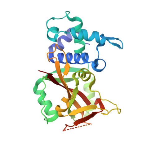
    6W65

  • PubMed Abstract: 

    Poly(ADP-ribose) polymerase (PARP) enzymes use nicotinamide adenine dinucleotide (NAD + ) to modify up to seven different amino acids with a single mono(ADP-ribose) unit (MARylation deposited by PARP monoenzymes) or branched poly(ADP-ribose) polymers (PARylation deposited by PARP polyenzymes). To enable the development of tool compounds for PARP monoenzymes and polyenzymes, we have developed active site probes for use in in vitro and cellular biophysical assays to characterize active site-directed inhibitors that compete for NAD + binding. These assays are agnostic of the protein substrate for each PARP, overcoming a general lack of knowledge around the substrates for these enzymes. The in vitro assays use less enzyme than previously described activity assays, enabling discrimination of inhibitor potencies in the single-digit nanomolar range, and the cell-based assays can differentiate compounds with sub-nanomolar potencies and measure inhibitor residence time in live cells.


  • Organizational Affiliation

    Ribon Therapeutics, 35 Cambridgepark Drive, Suite 300, Cambridge, MA 02140, USA. Electronic address: twigle@ribontx.com.


Macromolecules
Find similar proteins by:  (by identity cutoff)  |  3D Structure
Entity ID: 1
MoleculeChains Sequence LengthOrganismDetailsImage
Protein mono-ADP-ribosyltransferase PARP16
A, B, C
277Homo sapiensMutation(s): 0 
Gene Names: PARP16ARTD15C15orf30
EC: 2.4.2
UniProt & NIH Common Fund Data Resources
Find proteins for Q8N5Y8 (Homo sapiens)
Explore Q8N5Y8 
Go to UniProtKB:  Q8N5Y8
PHAROS:  Q8N5Y8
GTEx:  ENSG00000138617 
Entity Groups  
Sequence Clusters30% Identity50% Identity70% Identity90% Identity95% Identity100% Identity
UniProt GroupQ8N5Y8
Sequence Annotations
Expand
  • Reference Sequence
Small Molecules
Ligands 5 Unique
IDChains Name / Formula / InChI Key2D Diagram3D Interactions
T9D (Subject of Investigation/LOI)
Query on T9D

Download Ideal Coordinates CCD File 
D [auth A],
K [auth B],
U [auth C]
5-{5-[(piperidin-4-yl)oxy]-2H-isoindol-2-yl}-4-(trifluoromethyl)pyridazin-3(2H)-one
C18 H17 F3 N4 O2
DPAMCOKMVSYBNR-UHFFFAOYSA-N
CIT
Query on CIT

Download Ideal Coordinates CCD File 
E [auth A],
L [auth B]
CITRIC ACID
C6 H8 O7
KRKNYBCHXYNGOX-UHFFFAOYSA-N
PEG
Query on PEG

Download Ideal Coordinates CCD File 
R [auth B],
W [auth C]
DI(HYDROXYETHYL)ETHER
C4 H10 O3
MTHSVFCYNBDYFN-UHFFFAOYSA-N
GOL
Query on GOL

Download Ideal Coordinates CCD File 
F [auth A]
G [auth A]
H [auth A]
I [auth A]
M [auth B]
F [auth A],
G [auth A],
H [auth A],
I [auth A],
M [auth B],
N [auth B],
O [auth B],
P [auth B],
Q [auth B],
V [auth C]
GLYCEROL
C3 H8 O3
PEDCQBHIVMGVHV-UHFFFAOYSA-N
EDO
Query on EDO

Download Ideal Coordinates CCD File 
J [auth A],
S [auth B],
T [auth B],
X [auth C]
1,2-ETHANEDIOL
C2 H6 O2
LYCAIKOWRPUZTN-UHFFFAOYSA-N
Experimental Data & Validation

Experimental Data

  • Method: X-RAY DIFFRACTION
  • Resolution: 2.13 Å
  • R-Value Free: 0.225 
  • R-Value Work: 0.190 
  • R-Value Observed: 0.192 
  • Space Group: P 43 21 2
Unit Cell:
Length ( Å )Angle ( ˚ )
a = 147.12α = 90
b = 147.12β = 90
c = 99.44γ = 90
Software Package:
Software NamePurpose
REFMACrefinement
PDB_EXTRACTdata extraction
XDSdata reduction
XDSdata scaling
REFMACphasing

Structure Validation

View Full Validation Report


Ligand Structure Quality Assessment 


Entry History 

Deposition Data

Revision History  (Full details and data files)

  • Version 1.0: 2020-10-07
    Type: Initial release
  • Version 1.1: 2024-03-06
    Changes: Data collection, Database references, Refinement description