6UH8

Crystal structure of DAD2 N242I mutant


Experimental Data Snapshot

  • Method: X-RAY DIFFRACTION
  • Resolution: 1.58 Å
  • R-Value Free: 0.165 
  • R-Value Work: 0.138 
  • R-Value Observed: 0.140 

wwPDB Validation   3D Report Full Report


This is version 1.3 of the entry. See complete history


Literature

Flexibility of the petunia strigolactone receptor DAD2 promotes its interaction with signaling partners.

Lee, H.W.Sharma, P.Janssen, B.J.Drummond, R.S.M.Luo, Z.Hamiaux, C.Collier, T.Allison, J.R.Newcomb, R.D.Snowden, K.C.

(2020) J Biol Chem 295: 4181-4193

  • DOI: https://doi.org/10.1074/jbc.RA119.011509
  • Primary Citation of Related Structures:  
    6UH8, 6UH9

  • PubMed Abstract: 

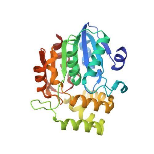
    Strigolactones (SLs) are terpenoid-derived plant hormones that regulate various developmental processes, particularly shoot branching, root development, and leaf senescence. The SL receptor has an unusual mode of action. Upon binding SL, it hydrolyzes the hormone, and then covalently binds one of the hydrolytic products. These initial events enable the SL receptor DAD2 (in petunia) to interact with the F-box protein PhMAX2A of the Skp-Cullin-F-box (SCF) complex and/or a repressor of SL signaling, PhD53A. However, it remains unclear how binding and hydrolysis structurally alters the SL receptor to enable its engagement with signaling partners. Here, we used mutagenesis to alter DAD2 and affect SL hydrolysis or DAD2's ability to interact with its signaling partners. We identified three DAD2 variants whose hydrolytic activity had been separated from the receptor's interactions with PhMAX2A or PhD53A. Two variants, DAD2 N242I and DAD2 F135A , having substitutions in the core α/β hydrolase-fold domain and the hairpin, exhibited hormone-independent interactions with PhMAX2A and PhD53A, respectively. Conversely, the DAD2 D166A variant could not interact with PhMAX2A in the presence of SL, but its interaction with PhD53A remained unaffected. Structural analyses of DAD2 N242I and DAD2 D166A revealed only small differences compared with the structure of the WT receptor. Results of molecular dynamics simulations of the DAD2 N242I structure suggested that increased flexibility is a likely cause for its SL-independent interaction with PhMAX2A. Our results suggest that PhMAX2A and PhD53A have distinct binding sites on the SL receptor and that its flexibility is a major determinant of its interactions with these two downstream regulators.


  • Organizational Affiliation

    The New Zealand Institute for Plant & Food Research Limited, Auckland, New Zealand; School of Biological Sciences, University of Auckland, Auckland, New Zealand.


Macromolecules
Find similar proteins by:  (by identity cutoff)  |  3D Structure
Entity ID: 1
MoleculeChains Sequence LengthOrganismDetailsImage
Decreased Apical Dominance 2
A, B
269Petunia x hybridaMutation(s): 2 
Gene Names: dad2
UniProt
Find proteins for J9U5U9 (Petunia hybrida)
Explore J9U5U9 
Go to UniProtKB:  J9U5U9
Entity Groups  
Sequence Clusters30% Identity50% Identity70% Identity90% Identity95% Identity100% Identity
UniProt GroupJ9U5U9
Sequence Annotations
Expand
  • Reference Sequence
Small Molecules
Ligands 4 Unique
IDChains Name / Formula / InChI Key2D Diagram3D Interactions
MES
Query on MES

Download Ideal Coordinates CCD File 
C [auth A],
F [auth B]
2-(N-MORPHOLINO)-ETHANESULFONIC ACID
C6 H13 N O4 S
SXGZJKUKBWWHRA-UHFFFAOYSA-N
SO4
Query on SO4

Download Ideal Coordinates CCD File 
M [auth B],
N [auth B],
O [auth B]
SULFATE ION
O4 S
QAOWNCQODCNURD-UHFFFAOYSA-L
GOL
Query on GOL

Download Ideal Coordinates CCD File 
E [auth A],
P [auth B]
GLYCEROL
C3 H8 O3
PEDCQBHIVMGVHV-UHFFFAOYSA-N
MG
Query on MG

Download Ideal Coordinates CCD File 
D [auth A]
G [auth B]
H [auth B]
I [auth B]
J [auth B]
D [auth A],
G [auth B],
H [auth B],
I [auth B],
J [auth B],
K [auth B],
L [auth B]
MAGNESIUM ION
Mg
JLVVSXFLKOJNIY-UHFFFAOYSA-N
Experimental Data & Validation

Experimental Data

  • Method: X-RAY DIFFRACTION
  • Resolution: 1.58 Å
  • R-Value Free: 0.165 
  • R-Value Work: 0.138 
  • R-Value Observed: 0.140 
  • Space Group: P 65
Unit Cell:
Length ( Å )Angle ( ˚ )
a = 133.736α = 90
b = 133.736β = 90
c = 99.764γ = 120
Software Package:
Software NamePurpose
XDSdata reduction
Aimlessdata scaling
PHASERphasing
REFMACrefinement
PDB_EXTRACTdata extraction

Structure Validation

View Full Validation Report


Entry History & Funding Information

Deposition Data


Funding OrganizationLocationGrant Number
Royal Society of New ZealandNew ZealandPAF1301

Revision History  (Full details and data files)

  • Version 1.0: 2020-02-26
    Type: Initial release
  • Version 1.1: 2020-03-04
    Changes: Database references
  • Version 1.2: 2020-04-08
    Changes: Database references
  • Version 1.3: 2023-10-11
    Changes: Data collection, Database references, Derived calculations, Refinement description