6SO5

Homo sapiens WRB/CAML heterotetramer in complex with a TRC40 dimer


Experimental Data Snapshot

  • Method: ELECTRON MICROSCOPY
  • Resolution: 4.20 Å
  • Aggregation State: PARTICLE 
  • Reconstruction Method: SINGLE PARTICLE 

wwPDB Validation   3D Report Full Report


This is version 1.1 of the entry. See complete history

Re-refinement Note

A newer entry is available that reflects an alternative modeling of the original data: 8CR1


Literature

Structural Basis of Tail-Anchored Membrane Protein Biogenesis by the GET Insertase Complex.

McDowell, M.A.Heimes, M.Fiorentino, F.Mehmood, S.Farkas, A.Coy-Vergara, J.Wu, D.Bolla, J.R.Schmid, V.Heinze, R.Wild, K.Flemming, D.Pfeffer, S.Schwappach, B.Robinson, C.V.Sinning, I.

(2020) Mol Cell 80: 72

  • DOI: https://doi.org/10.1016/j.molcel.2020.08.012
  • Primary Citation of Related Structures:  
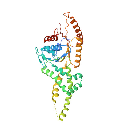
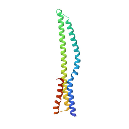
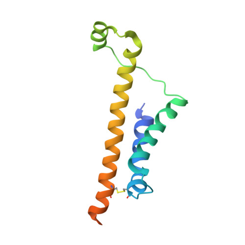
    6SO5

  • PubMed Abstract: 

    Membrane protein biogenesis faces the challenge of chaperoning hydrophobic transmembrane helices for faithful membrane insertion. The guided entry of tail-anchored proteins (GET) pathway targets and inserts tail-anchored (TA) proteins into the endoplasmic reticulum (ER) membrane with an insertase (yeast Get1/Get2 or mammalian WRB/CAML) that captures the TA from a cytoplasmic chaperone (Get3 or TRC40, respectively). Here, we present cryo-electron microscopy reconstructions, native mass spectrometry, and structure-based mutagenesis of human WRB/CAML/TRC40 and yeast Get1/Get2/Get3 complexes. Get3 binding to the membrane insertase supports heterotetramer formation, and phosphatidylinositol binding at the heterotetramer interface stabilizes the insertase for efficient TA insertion in vivo. We identify a Get2/CAML cytoplasmic helix that forms a "gating" interaction with Get3/TRC40 important for TA insertion. Structural homology with YidC and the ER membrane protein complex (EMC) implicates an evolutionarily conserved insertion mechanism for divergent substrates utilizing a hydrophilic groove. Thus, we provide a detailed structural and mechanistic framework to understand TA membrane insertion.


  • Organizational Affiliation

    Heidelberg University Biochemistry Center (BZH), Im Neuenheimer Feld 328, 69120 Heidelberg, Germany. Electronic address: melanie.mcdowell@bzh.uni-heidelberg.de.


Macromolecules
Find similar proteins by:  (by identity cutoff)  |  3D Structure
Entity ID: 1
MoleculeChains Sequence LengthOrganismDetailsImage
ATPase ASNA1
A, B
360Homo sapiensMutation(s): 0 
Gene Names: ASNA1ARSATRC40
EC: 3.6
UniProt & NIH Common Fund Data Resources
Find proteins for O43681 (Homo sapiens)
Explore O43681 
Go to UniProtKB:  O43681
PHAROS:  O43681
GTEx:  ENSG00000198356 
Entity Groups  
Sequence Clusters30% Identity50% Identity70% Identity90% Identity95% Identity100% Identity
UniProt GroupO43681
Sequence Annotations
Expand
  • Reference Sequence
Find similar proteins by:  (by identity cutoff)  |  3D Structure
Entity ID: 2
MoleculeChains Sequence LengthOrganismDetailsImage
Tail-anchored protein insertion receptor WRB
C, D
184Homo sapiensMutation(s): 0 
Gene Names: WRBCHD5
Membrane Entity: Yes 
UniProt & NIH Common Fund Data Resources
Find proteins for O00258 (Homo sapiens)
Explore O00258 
Go to UniProtKB:  O00258
PHAROS:  O00258
GTEx:  ENSG00000182093 
Entity Groups  
Sequence Clusters30% Identity50% Identity70% Identity90% Identity95% Identity100% Identity
UniProt GroupO00258
Sequence Annotations
Expand
  • Reference Sequence
Find similar proteins by:  (by identity cutoff)  |  3D Structure
Entity ID: 3
MoleculeChains Sequence LengthOrganismDetailsImage
Calcium signal-modulating cyclophilin ligand
E, F
127Homo sapiensMutation(s): 0 
Gene Names: CAMLGCAML
Membrane Entity: Yes 
UniProt & NIH Common Fund Data Resources
Find proteins for P49069 (Homo sapiens)
Explore P49069 
Go to UniProtKB:  P49069
PHAROS:  P49069
GTEx:  ENSG00000164615 
Entity Groups  
Sequence Clusters30% Identity50% Identity70% Identity90% Identity95% Identity100% Identity
UniProt GroupP49069
Sequence Annotations
Expand
  • Reference Sequence
Small Molecules
Ligands 1 Unique
IDChains Name / Formula / InChI Key2D Diagram3D Interactions
ZN
Query on ZN

Download Ideal Coordinates CCD File 
G [auth A]ZINC ION
Zn
PTFCDOFLOPIGGS-UHFFFAOYSA-N
Experimental Data & Validation

Experimental Data

  • Method: ELECTRON MICROSCOPY
  • Resolution: 4.20 Å
  • Aggregation State: PARTICLE 
  • Reconstruction Method: SINGLE PARTICLE 
EM Software:
TaskSoftware PackageVersion
RECONSTRUCTIONRELION3.0
MODEL REFINEMENTPHENIX

Structure Validation

View Full Validation Report


Entry History & Funding Information

Deposition Data


Funding OrganizationLocationGrant Number
German Research Foundation (DFG)GermanyLeibniz SI 586/6-1
German Research Foundation (DFG)GermanyTRR83 TP22
European Molecular Biology Organization (EMBO)GermanyALTF 1230-2013

Revision History  (Full details and data files)

  • Version 1.0: 2020-09-09
    Type: Initial release
  • Version 1.1: 2021-02-17
    Changes: Database references