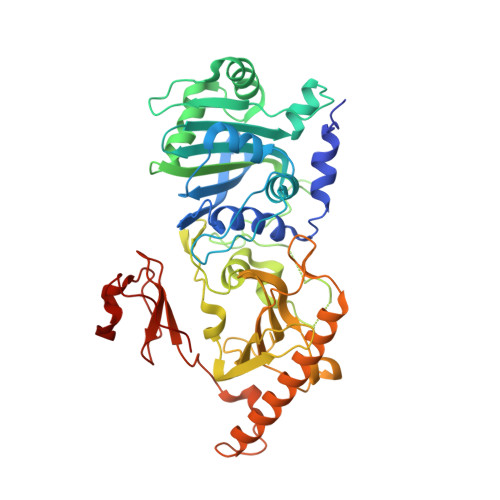
Mechanism for autoinhibition and activation of the MORC3 ATPase.
Zhang, Y., Klein, B.J., Cox, K.L., Bertulat, B., Tencer, A.H., Holden, M.R., Wright, G.M., Black, J., Cardoso, M.C., Poirier, M.G., Kutateladze, T.G.(2019) Proc Natl Acad Sci U S A 116: 6111-6119
- PubMed: 30850548
- DOI: https://doi.org/10.1073/pnas.1819524116
- Primary Citation of Related Structures:
6O1E - PubMed Abstract:
Microrchidia 3 (MORC3) is a human protein linked to autoimmune disorders, Down syndrome, and cancer. It is a member of a newly identified family of human ATPases with an uncharacterized mechanism of action. Here, we elucidate the molecular basis for inhibition and activation of MORC3. The crystal structure of the MORC3 region encompassing the ATPase and CW domains in complex with a nonhydrolyzable ATP analog demonstrates that the two domains are directly coupled. The extensive ATPase:CW interface stabilizes the protein fold but inhibits the catalytic activity of MORC3. Enzymatic, NMR, mutational, and biochemical analyses show that in the autoinhibited, off state, the CW domain sterically impedes binding of the ATPase domain to DNA, which in turn is required for the catalytic activity. MORC3 autoinhibition is released by disrupting the intramolecular ATPase:CW coupling through the competitive interaction of CW with histone H3 tail or by mutating the interfacial residues. Binding of CW to H3 leads to a marked rearrangement in the ATPase-CW cassette, which frees the DNA-binding site in active MORC3 (on state). We show that ATP-induced dimerization of the ATPase domain is strictly required for the catalytic activity and that the dimeric form of ATPase-CW might cooperatively bind to dsDNA. Together, our findings uncovered a mechanism underlying the fine-tuned regulation of the catalytic domain of MORC3 by the epigenetic reader, CW.
Organizational Affiliation:
Department of Pharmacology, University of Colorado School of Medicine, Aurora, CO 80045.