6ISP

structure of Candida antarctica Lipase B mutant


Experimental Data Snapshot

  • Method: X-RAY DIFFRACTION
  • Resolution: 1.88 Å
  • R-Value Free: 0.238 
  • R-Value Work: 0.213 
  • R-Value Observed: 0.215 

wwPDB Validation   3D Report Full Report


Ligand Structure Quality Assessment 


This is version 1.2 of the entry. See complete history


Literature

Artificial cysteine-lipases with high activity and altered catalytic mechanism created by laboratory evolution.

Cen, Y.Singh, W.Arkin, M.Moody, T.S.Huang, M.Zhou, J.Wu, Q.Reetz, M.T.

(2019) Nat Commun 10: 3198-3198

  • DOI: https://doi.org/10.1038/s41467-019-11155-3
  • Primary Citation of Related Structures:  
    6ISP, 6ISQ, 6ISR

  • PubMed Abstract: 

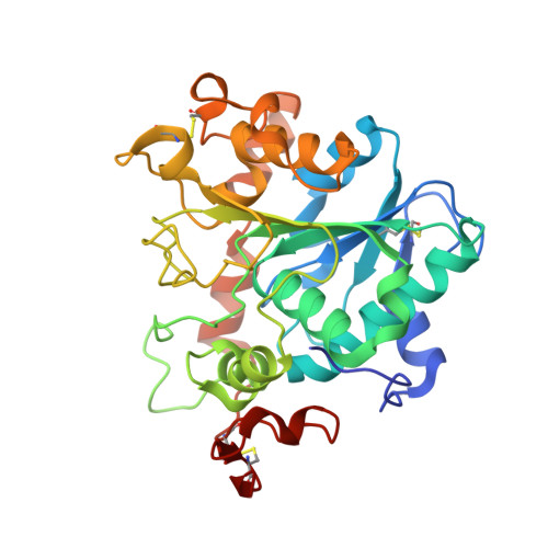
    Engineering artificial enzymes with high activity and catalytic mechanism different from naturally occurring enzymes is a challenge in protein design. For example, many attempts have been made to obtain active hydrolases by introducing a Ser → Cys exchange at the respective catalytic triads, but this generally induced a breakdown of activity. We now report that this long-standing dogma no longer pertains, provided additional mutations are introduced by directed evolution. By employing Candida antarctica lipase B (CALB) as the model enzyme with the Ser-His-Asp catalytic triad, a highly active cysteine-lipase having a Cys-His-Asp catalytic triad and additional mutations W104V/A281Y/A282Y/V149G can be evolved, showing a 40-fold higher catalytic efficiency than wild-type CALB in the hydrolysis of 4-nitrophenyl benzoate, and tolerating bulky substrates. Crystal structures, kinetics, MD simulations and QM/MM calculations reveal dynamic features and explain all results, including the preference of a two-step mechanism involving the zwitterionic pair Cys105 - /His224 + rather than a concerted process.


  • Organizational Affiliation

    Department of Chemistry, Zhejiang University, 310027, Hangzhou, China.


Macromolecules
Find similar proteins by:  (by identity cutoff)  |  3D Structure
Entity ID: 1
MoleculeChains Sequence LengthOrganismDetailsImage
Lipase BA [auth D],
B [auth A],
C,
D [auth B]
321Moesziomyces antarcticusMutation(s): 6 
EC: 3.1.1.3
Membrane Entity: Yes 
UniProt
Find proteins for P41365 (Pseudozyma antarctica)
Explore P41365 
Go to UniProtKB:  P41365
Entity Groups  
Sequence Clusters30% Identity50% Identity70% Identity90% Identity95% Identity100% Identity
UniProt GroupP41365
Sequence Annotations
Expand
  • Reference Sequence
Small Molecules
Ligands 2 Unique
IDChains Name / Formula / InChI Key2D Diagram3D Interactions
CPQ
Query on CPQ

Download Ideal Coordinates CCD File 
E [auth D]
F [auth D]
H [auth A]
I [auth A]
J [auth A]
E [auth D],
F [auth D],
H [auth A],
I [auth A],
J [auth A],
K [auth A],
L [auth C],
M [auth C],
N [auth C],
P [auth B],
Q [auth B]
N,N-BIS(3-D-GLUCONAMIDOPROPYL)DEOXYCHOLAMIDE
C42 H75 N3 O15
OJSUWTDDXLCUFR-HGZMBBKESA-N
CA
Query on CA

Download Ideal Coordinates CCD File 
G [auth A],
O [auth B]
CALCIUM ION
Ca
BHPQYMZQTOCNFJ-UHFFFAOYSA-N
Experimental Data & Validation

Experimental Data

  • Method: X-RAY DIFFRACTION
  • Resolution: 1.88 Å
  • R-Value Free: 0.238 
  • R-Value Work: 0.213 
  • R-Value Observed: 0.215 
  • Space Group: P 1 21 1
Unit Cell:
Length ( Å )Angle ( ˚ )
a = 46.98α = 90
b = 155.97β = 90
c = 92.53γ = 90
Software Package:
Software NamePurpose
PHENIXrefinement
XDSdata reduction
Aimlessdata scaling
PHENIXphasing

Structure Validation

View Full Validation Report


Ligand Structure Quality Assessment 


Entry History 

Deposition Data

Revision History  (Full details and data files)

  • Version 1.0: 2019-07-24
    Type: Initial release
  • Version 1.1: 2019-08-07
    Changes: Data collection, Database references
  • Version 1.2: 2023-11-22
    Changes: Data collection, Database references, Derived calculations, Refinement description