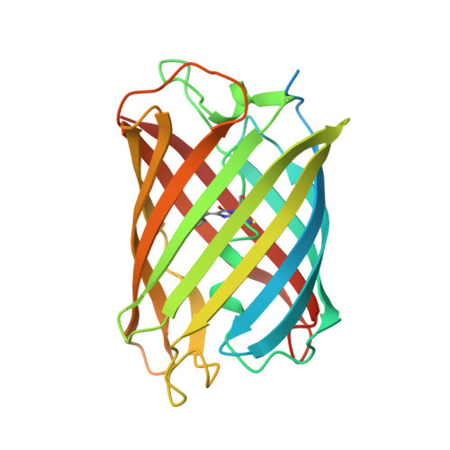
6D38

Photodissociable dimeric Dronpa green fluorescent protein variant M (pdDronpaM)


Experimental Data Snapshot

  • Method: X-RAY DIFFRACTION
  • Resolution: 2.80 Å
  • R-Value Free: 0.263 
  • R-Value Work: 0.195 
  • R-Value Observed: 0.199 

wwPDB Validation   3D Report Full Report


This is version 2.0 of the entry. See complete history


Literature

Optical control of cell signaling by single-chain photoswitchable kinases.

Zhou, X.X.Fan, L.Z.Li, P.Shen, K.Lin, M.Z.

(2017) Science 355: 836-842

  • DOI: https://doi.org/10.1126/science.aah3605
  • Primary Citation of Related Structures:  
    6D38, 6D39

  • PubMed Abstract: 

    Protein kinases transduce signals to regulate a wide array of cellular functions in eukaryotes. A generalizable method for optical control of kinases would enable fine spatiotemporal interrogation or manipulation of these various functions. We report the design and application of single-chain cofactor-free kinases with photoswitchable activity. We engineered a dimeric protein, pdDronpa, that dissociates in cyan light and reassociates in violet light. Attaching two pdDronpa domains at rationally selected locations in the kinase domain, we created the photoswitchable kinases psRaf1, psMEK1, psMEK2, and psCDK5. Using these photoswitchable kinases, we established an all-optical cell-based assay for screening inhibitors, uncovered a direct and rapid inhibitory feedback loop from ERK to MEK1, and mediated developmental changes and synaptic vesicle transport in vivo using light.


  • Organizational Affiliation

    Department of Bioengineering, Stanford University, Stanford, CA, USA.


Macromolecules
Find similar proteins by:  (by identity cutoff)  |  3D Structure
Entity ID: 1
MoleculeChains Sequence LengthOrganismDetailsImage
Fluorescent protein Dronpa259Echinophyllia sp. SC22Mutation(s): 5 
Gene Names: Dronpa
UniProt
Find proteins for Q5TLG6 (Echinophyllia sp. SC22)
Explore Q5TLG6 
Go to UniProtKB:  Q5TLG6
Entity Groups  
Sequence Clusters30% Identity50% Identity70% Identity90% Identity95% Identity100% Identity
UniProt GroupQ5TLG6
Sequence Annotations
Expand
  • Reference Sequence
Small Molecules
Modified Residues  1 Unique
IDChains TypeFormula2D DiagramParent
GYC
Query on GYC
A
L-PEPTIDE LINKINGC14 H15 N3 O4 SCYS, TYR, GLY
Experimental Data & Validation

Experimental Data

  • Method: X-RAY DIFFRACTION
  • Resolution: 2.80 Å
  • R-Value Free: 0.263 
  • R-Value Work: 0.195 
  • R-Value Observed: 0.199 
  • Space Group: P 43 21 2
Unit Cell:
Length ( Å )Angle ( ˚ )
a = 52.039α = 90
b = 52.039β = 90
c = 163.621γ = 90
Software Package:
Software NamePurpose
PHENIXrefinement
Cootmodel building
PDB_EXTRACTdata extraction
PHASERphasing
HKL-2000data scaling
HKL-2000data reduction

Structure Validation

View Full Validation Report


Entry History & Funding Information

Deposition Data


Funding OrganizationLocationGrant Number
National Institutes of Health/National Institute of General Medical Sciences (NIH/NIGMS)United States5DP1GM111003

Revision History  (Full details and data files)

  • Version 1.0: 2018-05-16
    Type: Initial release
  • Version 1.1: 2020-01-01
    Changes: Author supporting evidence
  • Version 1.2: 2023-10-04
    Changes: Data collection, Database references, Refinement description
  • Version 2.0: 2023-11-15
    Changes: Atomic model, Data collection, Derived calculations