6AEX

Crystal structure of unoccupied murine uPAR


Experimental Data Snapshot

  • Method: X-RAY DIFFRACTION
  • Resolution: 2.39 Å
  • R-Value Free: 0.244 
  • R-Value Work: 0.195 
  • R-Value Observed: 0.198 

wwPDB Validation   3D Report Full Report


Ligand Structure Quality Assessment 


This is version 1.2 of the entry. See complete history


Literature

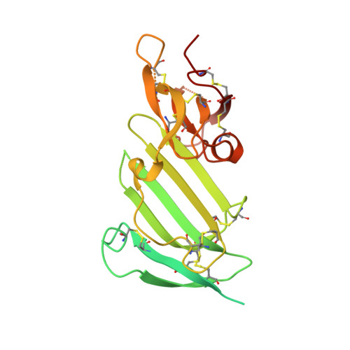
Crystal structure of the unoccupied murine urokinase-type plasminogen activator receptor (uPAR) reveals a tightly packed DII-DIII unit.

Liu, M.Lin, L.Hoyer-Hansen, G.Ploug, M.Li, H.Jiang, L.Yuan, C.Li, J.Huang, M.

(2019) FEBS Lett 593: 1236-1247

  • DOI: https://doi.org/10.1002/1873-3468.13397
  • Primary Citation of Related Structures:  
    6AEX

  • PubMed Abstract: 

    The urokinase-type plasminogen activator receptor (uPAR) is a cell surface receptor that is capable of binding to a range of extracellular proteins and triggering a series of proteolytic and signaling events. Previous structural studies of uPAR with its ligands uPA and vitronectin revealed that its three domains (DI, DII, and DIII) form a large hydrophobic cavity to accommodate uPA. In the present study, the structure of unoccupied murine uPAR (muPAR) is determined. The structure of DII and DIII of muPAR is well defined and forms a compact globular unit, while DI could not be traced. Molecular dynamic simulations further confirm the rigid binding interface between DII and DIII. This study shows overall structural flexibility of uPAR in the absence of uPA.


  • Organizational Affiliation

    College of Biological Science and Engineering, Fuzhou University, China.


Macromolecules
Find similar proteins by:  (by identity cutoff)  |  3D Structure
Entity ID: 1
MoleculeChains Sequence LengthOrganismDetailsImage
Urokinase plasminogen activator surface receptorA [auth U]277Mus musculusMutation(s): 0 
Gene Names: Plaur
UniProt & NIH Common Fund Data Resources
Find proteins for P35456 (Mus musculus)
Explore P35456 
Go to UniProtKB:  P35456
IMPC:  MGI:97612
Entity Groups  
Sequence Clusters30% Identity50% Identity70% Identity90% Identity95% Identity100% Identity
UniProt GroupP35456
Sequence Annotations
Expand
  • Reference Sequence
Small Molecules
Ligands 1 Unique
IDChains Name / Formula / InChI Key2D Diagram3D Interactions
NAG (Subject of Investigation/LOI)
Query on NAG

Download Ideal Coordinates CCD File 
B [auth U]2-acetamido-2-deoxy-beta-D-glucopyranose
C8 H15 N O6
OVRNDRQMDRJTHS-FMDGEEDCSA-N
Experimental Data & Validation

Experimental Data

  • Method: X-RAY DIFFRACTION
  • Resolution: 2.39 Å
  • R-Value Free: 0.244 
  • R-Value Work: 0.195 
  • R-Value Observed: 0.198 
  • Space Group: P 41 21 2
Unit Cell:
Length ( Å )Angle ( ˚ )
a = 83.82α = 90
b = 83.82β = 90
c = 81.398γ = 90
Software Package:
Software NamePurpose
PHENIXrefinement
HKL-2000data reduction
HKL-2000data scaling
MOLREPphasing

Structure Validation

View Full Validation Report


Ligand Structure Quality Assessment 


Entry History & Funding Information

Deposition Data


Funding OrganizationLocationGrant Number
National Natural Science Foundation of ChinaChina31370737
National Natural Science Foundation of ChinaChina31400637
National Natural Science Foundation of ChinaChina31570745
National Natural Science Foundation of ChinaChina31670739

Revision History  (Full details and data files)

  • Version 1.0: 2019-07-17
    Type: Initial release
  • Version 1.1: 2020-07-29
    Type: Remediation
    Reason: Carbohydrate remediation
    Changes: Data collection, Derived calculations, Structure summary
  • Version 1.2: 2023-11-22
    Changes: Data collection, Database references, Refinement description, Structure summary