6J20

Crystal structure of the human NK1 substance P receptor


Experimental Data Snapshot

  • Method: X-RAY DIFFRACTION
  • Resolution: 2.70 Å
  • R-Value Free: 0.260 
  • R-Value Work: 0.233 
  • R-Value Observed: 0.234 

wwPDB Validation   3D Report Full Report


Ligand Structure Quality Assessment 


This is version 1.1 of the entry. See complete history


Literature

Human substance P receptor binding mode of the antagonist drug aprepitant by NMR and crystallography.

Chen, S.Lu, M.Liu, D.Yang, L.Yi, C.Ma, L.Zhang, H.Liu, Q.Frimurer, T.M.Wang, M.W.Schwartz, T.W.Stevens, R.C.Wu, B.Wuthrich, K.Zhao, Q.

(2019) Nat Commun 10: 638-638

  • DOI: https://doi.org/10.1038/s41467-019-08568-5
  • Primary Citation of Related Structures:  
    6J20, 6J21

  • PubMed Abstract: 

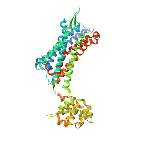
    Neurokinin 1 receptor (NK1R) has key regulating functions in the central and peripheral nervous systems, and NK1R antagonists such as aprepitant have been approved for treating chemotherapy-induced nausea and vomiting. However, the lack of data on NK1R structure and biochemistry has limited further drug development targeting this receptor. Here, we combine NMR spectroscopy and X-ray crystallography to provide dynamic and static characterisation of the binding mode of aprepitant in complexes with human NK1R variants. 19 F-NMR showed a slow off-rate in the binding site, where aprepitant occupies multiple substates that exchange with frequencies in the millisecond range. The environment of the bound ligand is affected by the amino acid in position 2.50, which plays a key role in ligand binding and receptor signaling in class A GPCRs. Crystal structures now reveal how receptor signaling relates to the conformation of the conserved NP 7.50 xxY motif in transmembrane helix VII.


  • Organizational Affiliation

    State Key Laboratory of Drug Research, Shanghai Institute of Materia Medica, Chinese Academy of Sciences, 555 Zuchongzhi Road, Pudong, Shanghai, 201203, China.


Macromolecules
Find similar proteins by:  (by identity cutoff)  |  3D Structure
Entity ID: 1
MoleculeChains Sequence LengthOrganismDetailsImage
Substance-P receptor,Endolysin441Homo sapiensTequatrovirus T4Mutation(s): 4 
Gene Names: TACR1NK1RTAC1ReT4Tp126
EC: 3.2.1.17
Membrane Entity: Yes 
UniProt & NIH Common Fund Data Resources
Find proteins for P00720 (Enterobacteria phage T4)
Explore P00720 
Go to UniProtKB:  P00720
Find proteins for P25103 (Homo sapiens)
Explore P25103 
Go to UniProtKB:  P25103
PHAROS:  P25103
GTEx:  ENSG00000115353 
Entity Groups  
Sequence Clusters30% Identity50% Identity70% Identity90% Identity95% Identity100% Identity
UniProt GroupsP00720P25103
Sequence Annotations
Expand
  • Reference Sequence
Small Molecules
Ligands 1 Unique
IDChains Name / Formula / InChI Key2D Diagram3D Interactions
GBQ
Query on GBQ

Download Ideal Coordinates CCD File 
B [auth A]5-[[(2~{R},3~{S})-2-[(1~{R})-1-[3,5-bis(trifluoromethyl)phenyl]ethoxy]-3-(4-fluorophenyl)morpholin-4-yl]methyl]-1,2-dihydro-1,2,4-triazol-3-one
C23 H21 F7 N4 O3
ATALOFNDEOCMKK-OITMNORJSA-N
Experimental Data & Validation

Experimental Data

  • Method: X-RAY DIFFRACTION
  • Resolution: 2.70 Å
  • R-Value Free: 0.260 
  • R-Value Work: 0.233 
  • R-Value Observed: 0.234 
  • Space Group: P 42 21 2
Unit Cell:
Length ( Å )Angle ( ˚ )
a = 102.512α = 90
b = 102.512β = 90
c = 156.962γ = 90
Software Package:
Software NamePurpose
BUSTERrefinement
XDSdata reduction
XSCALEdata scaling
PHASERphasing

Structure Validation

View Full Validation Report


Ligand Structure Quality Assessment 


Entry History & Funding Information

Deposition Data


Funding OrganizationLocationGrant Number
Chinese Academy of SciencesChinaQYZDB-SSW-SMC054

Revision History  (Full details and data files)

  • Version 1.0: 2019-03-06
    Type: Initial release
  • Version 1.1: 2023-11-22
    Changes: Data collection, Database references, Refinement description