6GV0

Insulin glulisine


Experimental Data Snapshot

  • Method: X-RAY DIFFRACTION
  • Resolution: 1.26 Å
  • R-Value Free: 0.152 
  • R-Value Work: 0.128 
  • R-Value Observed: 0.129 

wwPDB Validation   3D Report Full Report


This is version 1.2 of the entry. See complete history


Literature

Analysis of insulin glulisine at the molecular level by X-ray crystallography and biophysical techniques.

Gillis, R.B.Solomon, H.V.Govada, L.Oldham, N.J.Dinu, V.Jiwani, S.I.Gyasi-Antwi, P.Coffey, F.Meal, A.Morgan, P.S.Harding, S.E.Helliwell, J.R.Chayen, N.E.Adams, G.G.

(2021) Sci Rep 11: 1737-1737

  • DOI: https://doi.org/10.1038/s41598-021-81251-2
  • Primary Citation of Related Structures:  
    6GV0

  • PubMed Abstract: 

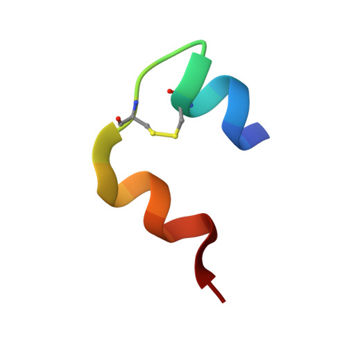
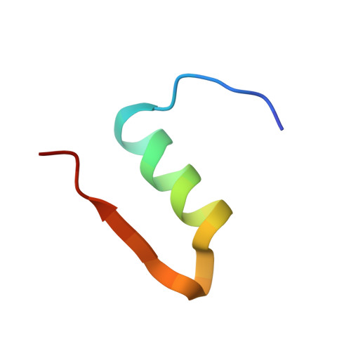
    This study concerns glulisine, a rapid-acting insulin analogue that plays a fundamental role in diabetes management. We have applied a combination of methods namely X-ray crystallography, and biophysical characterisation to provide a detailed insight into the structure and function of glulisine. X-ray data provided structural information to a resolution of 1.26 Å. Crystals belonged to the H3 space group with hexagonal (centred trigonal) cell dimensions a = b = 82.44 and c = 33.65 Å with two molecules in the asymmetric unit. A unique position of D21Glu, not present in other fast-acting analogues, pointing inwards rather than to the outside surface was observed. This reduces interactions with neighbouring molecules thereby increasing preference of the dimer form. Sedimentation velocity/equilibrium studies revealed a trinary system of dimers and hexamers/dihexamers in dynamic equilibrium. This new information may lead to better understanding of the pharmacokinetic and pharmacodynamic behaviour of glulisine which might aid in improving formulation regarding its fast-acting role and reducing side effects of this drug.


  • Organizational Affiliation

    Faculty of Medicine and Health Sciences, Queen's Medical Centre, University of Nottingham, Nottingham, NG7 2HA, UK. richard.gillis@nottingham.ac.uk.


Macromolecules

Find similar proteins by:  Sequence   |   3D Structure  

Entity ID: 1
MoleculeChains Sequence LengthOrganismDetailsImage
InsulinA [auth G],
C [auth I]
21Homo sapiensMutation(s): 0 
Gene Names: INS
UniProt & NIH Common Fund Data Resources
Find proteins for P01308 (Homo sapiens)
Explore P01308 
Go to UniProtKB:  P01308
PHAROS:  P01308
GTEx:  ENSG00000254647 
Entity Groups  
Sequence Clusters30% Identity50% Identity70% Identity90% Identity95% Identity100% Identity
UniProt GroupP01308
Sequence Annotations
Expand
  • Reference Sequence
Find similar proteins by:  (by identity cutoff)  |  3D Structure
Entity ID: 2
MoleculeChains Sequence LengthOrganismDetailsImage
Insulin
B, D
30Homo sapiensMutation(s): 2 
Gene Names: INS
UniProt & NIH Common Fund Data Resources
Find proteins for P01308 (Homo sapiens)
Explore P01308 
Go to UniProtKB:  P01308
PHAROS:  P01308
GTEx:  ENSG00000254647 
Entity Groups  
Sequence Clusters30% Identity50% Identity70% Identity90% Identity95% Identity100% Identity
UniProt GroupP01308
Sequence Annotations
Expand
  • Reference Sequence
Experimental Data & Validation

Experimental Data

Unit Cell:
Length ( Å )Angle ( ˚ )
a = 82.44α = 90
b = 82.44β = 90
c = 33.65γ = 120
Software Package:
Software NamePurpose
PHENIXrefinement
MOSFLMdata reduction
SCALAdata scaling
PHASERphasing

Structure Validation

View Full Validation Report


Entry History 

Deposition Data

Revision History  (Full details and data files)

  • Version 1.0: 2019-07-03
    Type: Initial release
  • Version 1.1: 2021-02-10
    Changes: Database references, Derived calculations
  • Version 1.2: 2024-01-17
    Changes: Data collection, Database references, Refinement description, Source and taxonomy