6E88

Cryo-EM structure of C. elegans GDP-microtubule


Experimental Data Snapshot

  • Method: ELECTRON MICROSCOPY
  • Resolution: 4.80 Å
  • Aggregation State: FILAMENT 
  • Reconstruction Method: SINGLE PARTICLE 

wwPDB Validation   3D Report Full Report


This is version 1.3 of the entry. See complete history


Literature

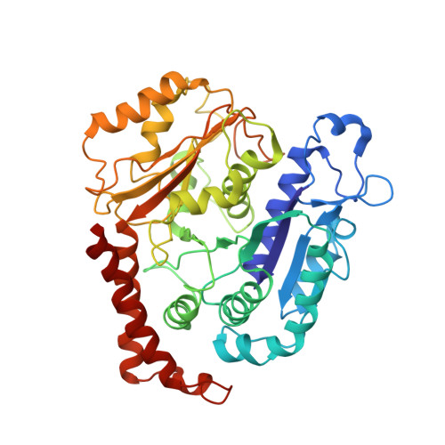
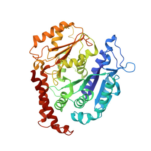
The Structure and Dynamics of C. elegans Tubulin Reveals the Mechanistic Basis of Microtubule Growth.

Chaaban, S.Jariwala, S.Hsu, C.T.Redemann, S.Kollman, J.M.Muller-Reichert, T.Sept, D.Bui, K.H.Brouhard, G.J.

(2018) Dev Cell 47: 191

  • DOI: https://doi.org/10.1016/j.devcel.2018.08.023
  • Primary Citation of Related Structures:  
    6E88

  • PubMed Abstract: 

    The dynamic instability of microtubules is a conserved and fundamental mechanism in eukaryotes. Yet microtubules from different species diverge in their growth rates, lattice structures, and responses to GTP hydrolysis. Therefore, we do not know what limits microtubule growth, what determines microtubule structure, or whether the mechanisms of dynamic instability are universal. Here, we studied microtubules from the nematode C. elegans, which have strikingly fast growth rates and non-canonical lattices in vivo. Using a reconstitution approach, we discovered that C. elegans microtubules combine intrinsically fast growth with very frequent catastrophes. We solved the structure of C. elegans microtubules to 4.8 Å and discovered sequence divergence in the lateral contact loops, one of which is ordered in C. elegans but unresolved in other species. We provide direct evidence that C. elegans tubulin has a higher free energy in solution and propose a model wherein the ordering of lateral contact loops activates tubulin for growth.


  • Organizational Affiliation

    Department of Biology, 1205 Avenue Docteur Penfield, Montréal, QC H3A 1B1, Canada.


Macromolecules
Find similar proteins by:  (by identity cutoff)  |  3D Structure
Entity ID: 1
MoleculeChains Sequence LengthOrganismDetailsImage
Tubulin alpha-2 chain434Caenorhabditis elegansMutation(s): 0 
UniProt
Find proteins for P34690 (Caenorhabditis elegans)
Explore P34690 
Go to UniProtKB:  P34690
Entity Groups  
Sequence Clusters30% Identity50% Identity70% Identity90% Identity95% Identity100% Identity
UniProt GroupP34690
Sequence Annotations
Expand
  • Reference Sequence
Find similar proteins by:  (by identity cutoff)  |  3D Structure
Entity ID: 2
MoleculeChains Sequence LengthOrganismDetailsImage
Tubulin beta-2 chain426Caenorhabditis elegansMutation(s): 0 
UniProt
Find proteins for P52275 (Caenorhabditis elegans)
Explore P52275 
Go to UniProtKB:  P52275
Entity Groups  
Sequence Clusters30% Identity50% Identity70% Identity90% Identity95% Identity100% Identity
UniProt GroupP52275
Sequence Annotations
Expand
  • Reference Sequence
Small Molecules
Ligands 2 Unique
IDChains Name / Formula / InChI Key2D Diagram3D Interactions
GTP
Query on GTP

Download Ideal Coordinates CCD File 
M [auth A]
O [auth C]
Q [auth H]
S [auth L]
U [auth I]
M [auth A],
O [auth C],
Q [auth H],
S [auth L],
U [auth I],
W [auth M]
GUANOSINE-5'-TRIPHOSPHATE
C10 H16 N5 O14 P3
XKMLYUALXHKNFT-UUOKFMHZSA-N
GDP
Query on GDP

Download Ideal Coordinates CCD File 
N [auth B]
P [auth D]
R [auth J]
T [auth N]
V [auth K]
N [auth B],
P [auth D],
R [auth J],
T [auth N],
V [auth K],
X [auth O]
GUANOSINE-5'-DIPHOSPHATE
C10 H15 N5 O11 P2
QGWNDRXFNXRZMB-UUOKFMHZSA-N
Experimental Data & Validation

Experimental Data

  • Method: ELECTRON MICROSCOPY
  • Resolution: 4.80 Å
  • Aggregation State: FILAMENT 
  • Reconstruction Method: SINGLE PARTICLE 

Structure Validation

View Full Validation Report


Entry History & Funding Information

Deposition Data


Funding OrganizationLocationGrant Number
Canadian Institutes of Health Research (CIHR)CanadaMOP-137055
Canadian Institutes of Health Research (CIHR)CanadaPJT-148702
Natural Sciences and Engineering Research Council (NSERC, Canada)Canada03791

Revision History  (Full details and data files)

  • Version 1.0: 2018-10-10
    Type: Initial release
  • Version 1.1: 2018-11-07
    Changes: Data collection, Database references, Structure summary
  • Version 1.2: 2020-01-15
    Changes: Author supporting evidence
  • Version 1.3: 2024-03-13
    Changes: Data collection, Database references