6DF3

Crystal structure of ternary complex of IL-24 with soluble receptors IL-22RA and IL-20RB


Experimental Data Snapshot

  • Method: X-RAY DIFFRACTION
  • Resolution: 2.15 Å
  • R-Value Free: 0.225 
  • R-Value Work: 0.182 
  • R-Value Observed: 0.184 

wwPDB Validation   3D Report Full Report


Ligand Structure Quality Assessment 


This is version 2.1 of the entry. See complete history


Literature

Crystal Structure of the Labile Complex of IL-24 with the Extracellular Domains of IL-22R1 and IL-20R2.

Lubkowski, J.Sonmez, C.Smirnov, S.V.Anishkin, A.Kotenko, S.V.Wlodawer, A.

(2018) J Immunol 201: 2082-2093

  • DOI: https://doi.org/10.4049/jimmunol.1800726
  • Primary Citation of Related Structures:  
    6DF3

  • PubMed Abstract: 

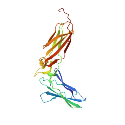
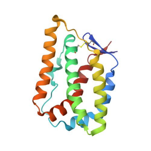
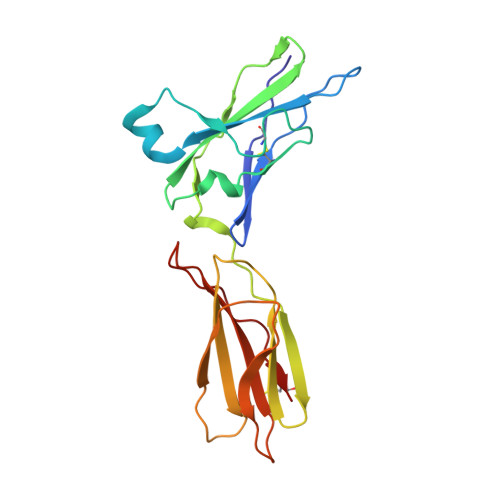
    Crystal structure of the ternary complex of human IL-24 with two receptors, IL-22R1 and IL-20R2, has been determined at 2.15 Å resolution. A crystallizable complex was created by a novel approach involving fusing the ligand with a flexible linker to the presumed low-affinity receptor, and coexpression of this construct in Drosophila S2 cells together with the presumed high-affinity receptor. This approach, which may be generally applicable to other multiprotein complexes with low-affinity components, was necessitated by the instability of IL-24 expressed by itself in either bacteria or insect cells. Although IL-24 expressed in Escherichia coli was unstable and precipitated almost immediately upon its refolding and purification, a small fraction of IL-24 remaining in the folded state was shown to be active in a cell-based assay. In the crystal structure presented here, we found that two cysteine residues in IL-24 do not form a predicted disulfide bond. Lack of structural restraint by disulfides, present in other related cytokines, is most likely reason for the low stability of IL-24. Although the contact area between IL-24 and IL-22R1 is larger than between the cytokine and IL-20R2, calculations show the latter interaction to be slightly more stable, suggesting that the shared receptor (IL-20R2) might be the higher-affinity receptor.


  • Organizational Affiliation

    Macromolecular Crystallography Laboratory, Center for Cancer Research, National Cancer Institute, Frederick, MD 21702; lubkowsj@mail.nih.gov.


Macromolecules
Find similar proteins by:  (by identity cutoff)  |  3D Structure
Entity ID: 1
MoleculeChains Sequence LengthOrganismDetailsImage
Interleukin-22 receptor subunit alpha-1A [auth L]206Homo sapiensMutation(s): 0 
Gene Names: IL22RA1IL22R
UniProt & NIH Common Fund Data Resources
Find proteins for Q8N6P7 (Homo sapiens)
Explore Q8N6P7 
Go to UniProtKB:  Q8N6P7
PHAROS:  Q8N6P7
GTEx:  ENSG00000142677 
Entity Groups  
Sequence Clusters30% Identity50% Identity70% Identity90% Identity95% Identity100% Identity
UniProt GroupQ8N6P7
Sequence Annotations
Expand
  • Reference Sequence
Find similar proteins by:  (by identity cutoff)  |  3D Structure
Entity ID: 2
MoleculeChains Sequence LengthOrganismDetailsImage
Interleukin-24B [auth C]155Homo sapiensMutation(s): 3 
Gene Names: IL24MDA7ST16
UniProt & NIH Common Fund Data Resources
Find proteins for Q13007 (Homo sapiens)
Explore Q13007 
Go to UniProtKB:  Q13007
PHAROS:  Q13007
GTEx:  ENSG00000162892 
Entity Groups  
Sequence Clusters30% Identity50% Identity70% Identity90% Identity95% Identity100% Identity
UniProt GroupQ13007
Sequence Annotations
Expand
  • Reference Sequence
Find similar proteins by:  (by identity cutoff)  |  3D Structure
Entity ID: 3
MoleculeChains Sequence LengthOrganismDetailsImage
Interleukin-20 receptor subunit betaC [auth H]190Homo sapiensMutation(s): 1 
Gene Names: IL20RBDIRS1UNQ557/PRO1114
UniProt & NIH Common Fund Data Resources
Find proteins for Q6UXL0 (Homo sapiens)
Explore Q6UXL0 
Go to UniProtKB:  Q6UXL0
PHAROS:  Q6UXL0
GTEx:  ENSG00000174564 
Entity Groups  
Sequence Clusters30% Identity50% Identity70% Identity90% Identity95% Identity100% Identity
UniProt GroupQ6UXL0
Sequence Annotations
Expand
  • Reference Sequence
Oligosaccharides

Help

Entity ID: 4
MoleculeChains Length2D Diagram Glycosylation3D Interactions
2-acetamido-2-deoxy-beta-D-glucopyranose-(1-4)-2-acetamido-2-deoxy-beta-D-glucopyranoseD [auth A]2N-Glycosylation
Glycosylation Resources
GlyTouCan:  G42666HT
GlyCosmos:  G42666HT
GlyGen:  G42666HT
Experimental Data & Validation

Experimental Data

  • Method: X-RAY DIFFRACTION
  • Resolution: 2.15 Å
  • R-Value Free: 0.225 
  • R-Value Work: 0.182 
  • R-Value Observed: 0.184 
  • Space Group: P 43
Unit Cell:
Length ( Å )Angle ( ˚ )
a = 77.702α = 90
b = 77.702β = 90
c = 124.785γ = 90
Software Package:
Software NamePurpose
HKL-3000data reduction
HKL-3000data scaling
PHASERphasing
REFMACrefinement
PDB_EXTRACTdata extraction

Structure Validation

View Full Validation Report


Ligand Structure Quality Assessment 


Entry History 

Deposition Data

Revision History  (Full details and data files)

  • Version 1.0: 2018-08-15
    Type: Initial release
  • Version 1.1: 2018-08-29
    Changes: Data collection, Database references
  • Version 1.2: 2018-10-03
    Changes: Data collection, Database references
  • Version 2.0: 2020-07-29
    Type: Remediation
    Reason: Carbohydrate remediation
    Changes: Atomic model, Data collection, Derived calculations, Structure summary
  • Version 2.1: 2023-10-11
    Changes: Data collection, Database references, Refinement description, Structure summary