5ZRF

Crystal structure of human topoisomerase II beta in complex with 5-iodouridine-containing-DNA and etoposide in space group p21


Experimental Data Snapshot

  • Method: X-RAY DIFFRACTION
  • Resolution: 2.30 Å
  • R-Value Free: 0.227 
  • R-Value Work: 0.189 
  • R-Value Observed: 0.189 

wwPDB Validation   3D Report Full Report


Ligand Structure Quality Assessment 


This is version 1.2 of the entry. See complete history


Literature

Structural insights into the gating of DNA passage by the topoisomerase II DNA-gate.

Chen, S.F.Huang, N.L.Lin, J.H.Wu, C.C.Wang, Y.R.Yu, Y.J.Gilson, M.K.Chan, N.L.

(2018) Nat Commun 9: 3085-3085

  • DOI: https://doi.org/10.1038/s41467-018-05406-y
  • Primary Citation of Related Structures:  
    5ZEN, 5ZQF, 5ZRF

  • PubMed Abstract: 

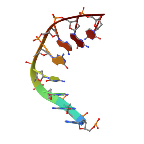
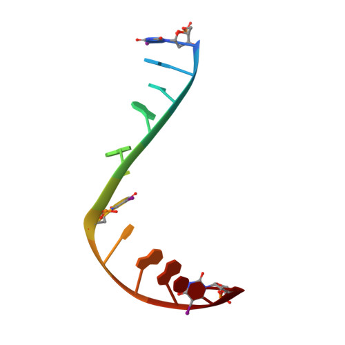
    Type IIA topoisomerases (Top2s) manipulate the handedness of DNA crossovers by introducing a transient and protein-linked double-strand break in one DNA duplex, termed the DNA-gate, whose opening allows another DNA segment to be transported through to change the DNA topology. Despite the central importance of this gate-opening event to Top2 function, the DNA-gate in all reported structures of Top2-DNA complexes is in the closed state. Here we present the crystal structure of a human Top2 DNA-gate in an open conformation, which not only reveals structural characteristics of its DNA-conducting path, but also uncovers unexpected yet functionally significant conformational changes associated with gate-opening. This structure further implicates Top2's preference for a left-handed DNA braid and allows the construction of a model representing the initial entry of another DNA duplex into the DNA-gate. Steered molecular dynamics calculations suggests the Top2-catalyzed DNA passage may be achieved by a rocker-switch-type movement of the DNA-gate.


  • Organizational Affiliation

    Institute of Biochemistry and Molecular Biology, College of Medicine, National Taiwan University, Taipei, 10051, Taiwan.


Macromolecules

Find similar proteins by:  (by identity cutoff)  |  3D Structure
Entity ID: 1
MoleculeChains Sequence LengthOrganismDetailsImage
DNA topoisomerase 2-beta
A, B
803Homo sapiensMutation(s): 0 
Gene Names: TOP2B
EC: 5.99.1.3
UniProt & NIH Common Fund Data Resources
Find proteins for Q02880 (Homo sapiens)
Explore Q02880 
Go to UniProtKB:  Q02880
PHAROS:  Q02880
GTEx:  ENSG00000077097 
Entity Groups  
Sequence Clusters30% Identity50% Identity70% Identity90% Identity95% Identity100% Identity
UniProt GroupQ02880
Sequence Annotations
Expand
  • Reference Sequence

Find similar nucleic acids by:  Sequence   |   3D Structure  

Entity ID: 2
MoleculeChains LengthOrganismImage
DNA (5'-D(P*AP*GP*CP*CP*GP*AP*GP*C)-3')
C, E
8synthetic construct
Sequence Annotations
Expand
  • Reference Sequence

Find similar nucleic acids by:  Sequence   |   3D Structure  

Entity ID: 3
MoleculeChains LengthOrganismImage
DNA/RNA (5'-R(P*(IU))-D(P*GP*CP*AP*GP*C)-R(P*(IU))-D(P*CP*GP*GP*C)-R(P*(IU))-3')
D, F
12synthetic construct
Sequence Annotations
Expand
  • Reference Sequence
Small Molecules
Ligands 2 Unique
IDChains Name / Formula / InChI Key2D Diagram3D Interactions
EVP
Query on EVP

Download Ideal Coordinates CCD File 
K [auth D],
M [auth F]
(5S,5aR,8aR,9R)-9-(4-hydroxy-3,5-dimethoxyphenyl)-8-oxo-5,5a,6,8,8a,9-hexahydrofuro[3',4':6,7]naphtho[2,3-d][1,3]dioxol -5-yl 4,6-O-[(1R)-ethylidene]-beta-D-glucopyranoside
C29 H32 O13
VJJPUSNTGOMMGY-MRVIYFEKSA-N
MG
Query on MG

Download Ideal Coordinates CCD File 
G [auth A]
H [auth A]
I [auth B]
J [auth B]
L [auth D]
G [auth A],
H [auth A],
I [auth B],
J [auth B],
L [auth D],
N [auth F]
MAGNESIUM ION
Mg
JLVVSXFLKOJNIY-UHFFFAOYSA-N
Binding Affinity Annotations 
IDSourceBinding Affinity
EVP BindingDB:  5ZRF IC50: min: 70, max: 790 (nM) from 2 assay(s)
EC50: 7.00e+4 (nM) from 1 assay(s)
Experimental Data & Validation

Experimental Data

  • Method: X-RAY DIFFRACTION
  • Resolution: 2.30 Å
  • R-Value Free: 0.227 
  • R-Value Work: 0.189 
  • R-Value Observed: 0.189 
  • Space Group: P 1 21 1
Unit Cell:
Length ( Å )Angle ( ˚ )
a = 80.147α = 90
b = 176.27β = 111.44
c = 94.331γ = 90
Software Package:
Software NamePurpose
PHENIXrefinement
HKL-2000data collection
HKL-2000data scaling
PHENIXphasing

Structure Validation

View Full Validation Report


Ligand Structure Quality Assessment 


Entry History 

Deposition Data

Revision History  (Full details and data files)

  • Version 1.0: 2018-08-08
    Type: Initial release
  • Version 1.1: 2019-02-20
    Changes: Data collection, Database references
  • Version 1.2: 2023-11-22
    Changes: Data collection, Database references, Derived calculations, Refinement description, Structure summary