5Z3S

Crystal structure of butanol modified signaling protein from buffalo (SPB-40) at 1.65 A resolution


Experimental Data Snapshot

  • Method: X-RAY DIFFRACTION
  • Resolution: 1.65 Å
  • R-Value Free: 0.206 
  • R-Value Work: 0.165 
  • R-Value Observed: 0.167 

wwPDB Validation   3D Report Full Report


Ligand Structure Quality Assessment 


This is version 1.4 of the entry. See complete history


Literature

A glycoprotein from mammary gland secreted during involution promotes apoptosis: Structural and biological studies.

Chaudhary, A.Kumar, V.Singh, P.K.Sharma, P.Bairagya, H.R.Kaur, P.Sharma, S.Chauhan, S.S.Singh, T.P.

(2018) Arch Biochem Biophys 644: 72-80

  • DOI: https://doi.org/10.1016/j.abb.2018.03.006
  • Primary Citation of Related Structures:  
    5Z05, 5Z3S, 5Z4W

  • PubMed Abstract: 

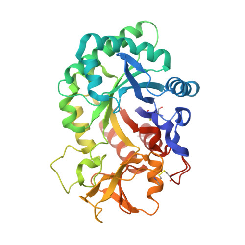
    Secretory signalling glycoprotein (SPX-40) from mammary gland is highly expressed during involution. This protein is involved in a programmed cell death during tissue remodelling which occurs at the end of lactation. SPX-40 was isolated and purified from buffalo (SPB-40) from the samples obtained during involution. One solution of SPB-40 was made by dissolving it in buffer containing 25 mM Tris-HCl and 50 mM NaCl at pH 8.0. Another solution was made by adding 25% ethanol to the above solution. The biological effects of SPB-40 dissolved in above two solutions were evaluated on MCF-7 breast cancer cell lines. Free SPB-40 indicated significant pro-apoptotic effects while ethanol exposed SPB-40 showed considerably reduced effects on the apoptosis. SPB-40 was crystallized in the native state. The crystals of SPB-40 were soaked in four separate solutions containing 25% acetone, 25% ethanol, 25% butanol and 25% MPD. Four separate data sets were collected and their structures were determined at high resolutions. In all the four structures, the molecules of acetone, ethanol, butanol and MPD respectively were observed in the hydrophobic binding pocket of SPB-40. As a result of which, the conformation of Trp78 was altered thus blocking the binding site in SPB-40 leading to the loss of activity.


  • Organizational Affiliation

    Department of Biophysics, All India Institute of Medical Sciences, New Delhi, India.


Macromolecules
Find similar proteins by:  (by identity cutoff)  |  3D Structure
Entity ID: 1
MoleculeChains Sequence LengthOrganismDetailsImage
Chitinase-3-like protein 1361Bubalus bubalisMutation(s): 0 
UniProt
Find proteins for Q7YS85 (Bubalus bubalis)
Explore Q7YS85 
Go to UniProtKB:  Q7YS85
Entity Groups  
Sequence Clusters30% Identity50% Identity70% Identity90% Identity95% Identity100% Identity
UniProt GroupQ7YS85
Sequence Annotations
Expand
  • Reference Sequence
Small Molecules
Experimental Data & Validation

Experimental Data

  • Method: X-RAY DIFFRACTION
  • Resolution: 1.65 Å
  • R-Value Free: 0.206 
  • R-Value Work: 0.165 
  • R-Value Observed: 0.167 
  • Space Group: P 21 21 21
Unit Cell:
Length ( Å )Angle ( ˚ )
a = 61.142α = 90
b = 66.553β = 90
c = 106.005γ = 90
Software Package:
Software NamePurpose
REFMACrefinement
HKL-2000data reduction
SCALEPACKdata scaling
MOLREPphasing

Structure Validation

View Full Validation Report


Ligand Structure Quality Assessment 


Entry History 

Deposition Data

Revision History  (Full details and data files)

  • Version 1.0: 2018-02-14
    Type: Initial release
  • Version 1.1: 2018-03-21
    Changes: Database references
  • Version 1.2: 2018-03-28
    Changes: Database references
  • Version 1.3: 2020-07-29
    Type: Remediation
    Reason: Carbohydrate remediation
    Changes: Data collection, Derived calculations, Structure summary
  • Version 1.4: 2023-11-22
    Changes: Data collection, Database references, Refinement description, Structure summary