5WV3

Crystal structure of bovine lactoperoxidase with a partial Glu258-heme linkage at 2.07 A resolution.


Experimental Data Snapshot

  • Method: X-RAY DIFFRACTION
  • Resolution: 2.07 Å
  • R-Value Free: 0.236 
  • R-Value Work: 0.167 
  • R-Value Observed: 0.169 

wwPDB Validation   3D Report Full Report


Ligand Structure Quality Assessment 


This is version 2.1 of the entry. See complete history

Re-refinement Note

This entry reflects an alternative modeling of the original data in: 5K1E


Literature

Structural basis of activation of mammalian heme peroxidases

Singh, P.K.Iqbal, N.Sirohi, H.V.Bairagya, H.R.Kaur, P.Sharma, S.Singh, T.P.

(2018) Prog Biophys Mol Biol 133: 49-55

  • DOI: https://doi.org/10.1016/j.pbiomolbio.2017.11.003
  • Primary Citation of Related Structures:  
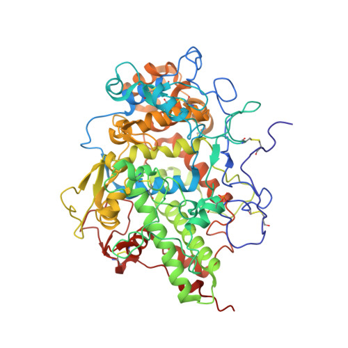
    5WV3

  • PubMed Abstract: 

    The mammalian heme peroxidases including lactoperoxidase (LPO), myeloperoxidase (MPO), eosinophil peroxidase (EPO) and thyroid peroxidase (TPO) contain a covalently linked heme moiety. Initially, it was believed that the heme group was fully cross-linked to protein molecule through at least two ester linkages involving conserved glutamate and aspartate residues with 1-methyl and 5-methyl groups of pyrrole rings A and C respectively. In MPO, an additional sulfonium ion linkage was present between 2-vinyl group of pyrrole ring A of the heme moiety and a methionine residue of the protein. These linkages were formed through a self processing mechanism. Subsequently, biochemical studies indicated that the heme moiety was partially attached to protein. The recent structural studies have shown that the covalent linkage involving glutamate and 1-methyl group of pyrrole ring of heme moiety was partially formed. When glutamate is not covalently linked to heme moiety, its side chain occupies a position in the substrate binding site on the distal heme side and blocks the substrate binding site leading to inactivation. However, an exposure to H 2 O 2 converts it to a fully covalently linked state with heme. Thus in mammalian heme peroxidases, the Glu-heme linkage is essential for catalytic action.


  • Organizational Affiliation

    Department of Biophysics, All India Institute of Medical Sciences, New Delhi, India.


Macromolecules
Find similar proteins by:  (by identity cutoff)  |  3D Structure
Entity ID: 1
MoleculeChains Sequence LengthOrganismDetailsImage
Lactoperoxidase595Bos taurusMutation(s): 0 
EC: 1.11.1.7
UniProt
Find proteins for P80025 (Bos taurus)
Explore P80025 
Go to UniProtKB:  P80025
Entity Groups  
Sequence Clusters30% Identity50% Identity70% Identity90% Identity95% Identity100% Identity
UniProt GroupP80025
Sequence Annotations
Expand
  • Reference Sequence
Oligosaccharides

Help

Entity ID: 2
MoleculeChains Length2D Diagram Glycosylation3D Interactions
2-acetamido-2-deoxy-beta-D-glucopyranose-(1-4)-2-acetamido-2-deoxy-beta-D-glucopyranose
B
2N-Glycosylation
Glycosylation Resources
GlyTouCan:  G42666HT
GlyCosmos:  G42666HT
GlyGen:  G42666HT
Small Molecules
Ligands 7 Unique
IDChains Name / Formula / InChI Key2D Diagram3D Interactions
HEM
Query on HEM

Download Ideal Coordinates CCD File 
D [auth A]PROTOPORPHYRIN IX CONTAINING FE
C34 H32 Fe N4 O4
KABFMIBPWCXCRK-RGGAHWMASA-L
NAG
Query on NAG

Download Ideal Coordinates CCD File 
E [auth A],
F [auth A],
G [auth A]
2-acetamido-2-deoxy-beta-D-glucopyranose
C8 H15 N O6
OVRNDRQMDRJTHS-FMDGEEDCSA-N
IOD
Query on IOD

Download Ideal Coordinates CCD File 
H [auth A],
S [auth A],
U [auth A]
IODIDE ION
I
XMBWDFGMSWQBCA-UHFFFAOYSA-M
GOL
Query on GOL

Download Ideal Coordinates CCD File 
R [auth A],
W [auth A]
GLYCEROL
C3 H8 O3
PEDCQBHIVMGVHV-UHFFFAOYSA-N
BR
Query on BR

Download Ideal Coordinates CCD File 
T [auth A]BROMIDE ION
Br
CPELXLSAUQHCOX-UHFFFAOYSA-M
OSM
Query on OSM

Download Ideal Coordinates CCD File 
I [auth A]
J [auth A]
K [auth A]
L [auth A]
M [auth A]
I [auth A],
J [auth A],
K [auth A],
L [auth A],
M [auth A],
N [auth A],
O [auth A],
P [auth A],
Q [auth A],
V [auth A]
1-(OXIDOSULFANYL)METHANAMINE
C H5 N O S
KONFWJXIYYGXOO-UHFFFAOYSA-N
CA
Query on CA

Download Ideal Coordinates CCD File 
C [auth A]CALCIUM ION
Ca
BHPQYMZQTOCNFJ-UHFFFAOYSA-N
Modified Residues  1 Unique
IDChains TypeFormula2D DiagramParent
SEP
Query on SEP
A
L-PEPTIDE LINKINGC3 H8 N O6 PSER
Experimental Data & Validation

Experimental Data

  • Method: X-RAY DIFFRACTION
  • Resolution: 2.07 Å
  • R-Value Free: 0.236 
  • R-Value Work: 0.167 
  • R-Value Observed: 0.169 
  • Space Group: P 1 21 1
Unit Cell:
Length ( Å )Angle ( ˚ )
a = 54.012α = 90
b = 79.809β = 93.01
c = 66.106γ = 90
Software Package:
Software NamePurpose
REFMACrefinement
HKL-2000data reduction
SCALEPACKdata scaling
MOLREPphasing

Structure Validation

View Full Validation Report


Ligand Structure Quality Assessment 


Entry History 

Deposition Data

Revision History  (Full details and data files)

  • Version 1.0: 2017-02-15
    Type: Initial release
  • Version 1.1: 2017-03-08
    Changes: Other
  • Version 1.2: 2018-04-25
    Changes: Data collection, Database references
  • Version 1.3: 2018-07-18
    Changes: Data collection, Database references
  • Version 2.0: 2020-07-29
    Type: Remediation
    Reason: Carbohydrate remediation
    Changes: Atomic model, Data collection, Derived calculations, Structure summary
  • Version 2.1: 2023-11-22
    Changes: Data collection, Database references, Derived calculations, Refinement description, Structure summary