5W8O

Homoserine transacetylase MetX from Mycobacterium hassiacum


Experimental Data Snapshot

  • Method: X-RAY DIFFRACTION
  • Resolution: 1.47 Å
  • R-Value Free: 0.183 
  • R-Value Work: 0.153 
  • R-Value Observed: 0.154 

wwPDB Validation   3D Report Full Report


This is version 1.5 of the entry. See complete history


Literature

Structural analysis of mycobacterial homoserine transacetylases central to methionine biosynthesis reveals druggable active site.

Chaton, C.T.Rodriguez, E.S.Reed, R.W.Li, J.Kenner, C.W.Korotkov, K.V.

(2019) Sci Rep 9: 20267-20267

  • DOI: https://doi.org/10.1038/s41598-019-56722-2
  • Primary Citation of Related Structures:  
    5W8O, 5W8P, 6PUX

  • PubMed Abstract: 

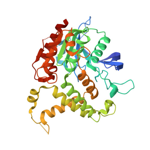
    Mycobacterium tuberculosis is the cause of the world's most deadly infectious disease. Efforts are underway to target the methionine biosynthesis pathway, as it is not part of the host metabolism. The homoserine transacetylase MetX converts L-homoserine to O-acetyl-L-homoserine at the committed step of this pathway. In order to facilitate structure-based drug design, we determined the high-resolution crystal structures of three MetX proteins, including M. tuberculosis (MtMetX), Mycolicibacterium abscessus (MaMetX), and Mycolicibacterium hassiacum (MhMetX). A comparison of homoserine transacetylases from other bacterial and fungal species reveals a high degree of structural conservation amongst the enzymes. Utilizing homologous structures with bound cofactors, we analyzed the potential ligandability of MetX. The deep active-site tunnel surrounding the catalytic serine yielded many consensus clusters during mapping, suggesting that MtMetX is highly druggable.


  • Organizational Affiliation

    Department of Molecular & Cellular Biochemistry and the Center for Structural Biology, University of Kentucky, Lexington, KY, 40536, USA.


Macromolecules
Find similar proteins by:  (by identity cutoff)  |  3D Structure
Entity ID: 1
MoleculeChains Sequence LengthOrganismDetailsImage
Homoserine O-acetyltransferase
A, B
363Mycolicibacterium hassiacum DSM 44199Mutation(s): 0 
Gene Names: metXC731_1248
EC: 2.3.1.31
UniProt
Find proteins for K5B926 (Mycolicibacterium hassiacum (strain DSM 44199 / CIP 105218 / JCM 12690 / 3849))
Explore K5B926 
Go to UniProtKB:  K5B926
Entity Groups  
Sequence Clusters30% Identity50% Identity70% Identity90% Identity95% Identity100% Identity
UniProt GroupK5B926
Sequence Annotations
Expand
  • Reference Sequence
Experimental Data & Validation

Experimental Data

  • Method: X-RAY DIFFRACTION
  • Resolution: 1.47 Å
  • R-Value Free: 0.183 
  • R-Value Work: 0.153 
  • R-Value Observed: 0.154 
  • Space Group: P 1 21 1
Unit Cell:
Length ( Å )Angle ( ˚ )
a = 47.75α = 90
b = 93.23β = 94.78
c = 81.59γ = 90
Software Package:
Software NamePurpose
XSCALEdata scaling
PHASERphasing
PHENIXrefinement
PDB_EXTRACTdata extraction
XDSdata reduction

Structure Validation

View Full Validation Report


Entry History & Funding Information

Deposition Data


Funding OrganizationLocationGrant Number
National Institutes of Health/National Institute of General Medical Sciences (NIH/NIGMS)United StatesP20GM103486
National Institutes of Health/National Institute of General Medical Sciences (NIH/NIGMS)United StatesP30GM110787

Revision History  (Full details and data files)

  • Version 1.0: 2017-07-12
    Type: Initial release
  • Version 1.1: 2017-09-27
    Changes: Author supporting evidence
  • Version 1.2: 2019-11-27
    Changes: Database references, Structure summary
  • Version 1.3: 2020-01-01
    Changes: Author supporting evidence
  • Version 1.4: 2020-01-15
    Changes: Database references
  • Version 1.5: 2023-10-04
    Changes: Data collection, Database references, Derived calculations, Refinement description