NMR structures in different membrane environments of three ocellatin peptides isolated from Leptodactylus labyrinthicus.
Gomes, K.A.G.G., Dos Santos, D.M., Santos, V.M., Pilo-Veloso, D., Mundim, H.M., Rodrigues, L.V., Liao, L.M., Verly, R.M., de Lima, M.E., Resende, J.M.(2018) Peptides 103: 72-83
- PubMed: 29596881
- DOI: https://doi.org/10.1016/j.peptides.2018.03.016
- Primary Citation of Related Structures:
5U9Q, 5U9R, 5U9Y, 5UA6, 5UA7, 5UA8 - PubMed Abstract:
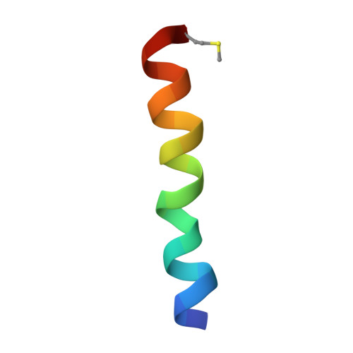
The peptides ocellatin-LB1, -LB2 and -F1 have previously been isolated from anurans of the Leptodactylus genus and the sequences are identical from residue 1-22, which correspond to ocellatin-LB1 sequence (GVVDILKGAAKDIAGHLASKVM-NH 2 ), whereas ocellatin-LB2 carries an extra N and ocellatin-F1 extra NKL residues at their C-termini. These peptides showed different spectra of activities and biophysical investigations indicated a direct correlation between membrane-disruptive properties and antimicrobial activities, i.e. ocellatin-F1 > ocellatin-LB1 > ocellatin-LB2. To better characterize their membrane interactions, we report here the detailed three-dimensional NMR structures of these peptides in TFE-d 2 :H 2 O (60:40) and in the presence of zwitterionic DPC-d 38 and anionic SDS-d 25 micellar solutions. Although the three peptides showed significant helical contents in the three mimetic environments, structural differences were noticed. When the structures of the three peptides in the presence of DPC-d 38 micelles are compared to each other, a more pronounced curvature is observed for ocellatin-F1 and the bent helix, with the concave face composed mostly of hydrophobic residues, is consistent with the micellar curvature and the amphipathic nature of the molecule. Interestingly, an almost linear helical segment was observed for ocellatin-F1 in the presence of SDS-d 25 micelles and the conformational differences in the two micellar environments are possibly related to the presence of the extra Lys residue near the peptide C-terminus, which increases the affinity of ocellatin-F1 to anionic membranes in comparison with ocellatin-LB1 and -LB2, as proved by isothermal titration calorimetry. To our knowledge, this work reports for the first time the three-dimensional structures of ocellatin peptides.
- Departamento de Química, Instituto de Ciências Exatas, Universidade Federal de Minas Gerais, P.O. Box 486, 31270-901 Belo Horizonte, MG, Brazil; Instituto de Engenharia, Ciência e Tecnologia, Universidade Federal dos Vales do Jequitinhonha e Mucuri, 39440-000 Janaúba, MG, Brazil.
Organizational Affiliation: