5SUY

Domain-swapped dimer of human Dishevelled2 DEP domain: monoclinic crystal form crystallised from dimeric fraction


Experimental Data Snapshot

  • Method: X-RAY DIFFRACTION
  • Resolution: 1.88 Å
  • R-Value Free: 0.217 
  • R-Value Work: 0.185 
  • R-Value Observed: 0.186 

wwPDB Validation   3D Report Full Report


This is version 1.2 of the entry. See complete history


Literature

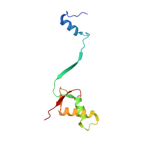
Wnt Signalosome Assembly by DEP Domain Swapping of Dishevelled.

Gammons, M.V.Renko, M.Johnson, C.M.Rutherford, T.J.Bienz, M.

(2016) Mol Cell 64: 92-104

  • DOI: https://doi.org/10.1016/j.molcel.2016.08.026
  • Primary Citation of Related Structures:  
    5LNP, 5SUY, 5SUZ

  • PubMed Abstract: 

    Extracellular signals are often transduced by dynamic signaling complexes ("signalosomes") assembled by oligomerizing hub proteins following their recruitment to signal-activated transmembrane receptors. A paradigm is the Wnt signalosome, which is assembled by Dishevelled via reversible head-to-tail polymerization by its DIX domain. Its activity causes stabilization of β-catenin, a Wnt effector with pivotal roles in animal development and cancer. How Wnt triggers signalosome assembly is unknown. Here, we use structural analysis, as well as biophysical and cell-based assays, to show that the DEP domain of Dishevelled undergoes a conformational switch, from monomeric to swapped dimer, to trigger DIX-dependent polymerization and signaling to β-catenin. This occurs in two steps: binding of monomeric DEP to Frizzled followed by DEP domain swapping triggered by its high local concentration upon Wnt-induced recruitment into clathrin-coated pits. DEP domain swapping confers directional bias on signaling, and the dimerization provides cross-linking between Dishevelled polymers, illustrating a key principle underlying signalosome formation.


  • Organizational Affiliation

    MRC Laboratory of Molecular Biology, Cambridge Biomedical Campus, Francis Crick Avenue, Cambridge CB2 0QH, UK. Electronic address: melissag@mrc-lmb.cam.ac.uk.


Macromolecules
Find similar proteins by:  (by identity cutoff)  |  3D Structure
Entity ID: 1
MoleculeChains Sequence LengthOrganismDetailsImage
Segment polarity protein dishevelled homolog DVL-2
A, B, C, D
97Homo sapiensMutation(s): 0 
Gene Names: DVL2
UniProt & NIH Common Fund Data Resources
Find proteins for O14641 (Homo sapiens)
Explore O14641 
Go to UniProtKB:  O14641
PHAROS:  O14641
GTEx:  ENSG00000004975 
Entity Groups  
Sequence Clusters30% Identity50% Identity70% Identity90% Identity95% Identity100% Identity
UniProt GroupO14641
Sequence Annotations
Expand
  • Reference Sequence
Experimental Data & Validation

Experimental Data

  • Method: X-RAY DIFFRACTION
  • Resolution: 1.88 Å
  • R-Value Free: 0.217 
  • R-Value Work: 0.185 
  • R-Value Observed: 0.186 
  • Space Group: P 1 21 1
Unit Cell:
Length ( Å )Angle ( ˚ )
a = 61.671α = 90
b = 60.174β = 117.62
c = 66.128γ = 90
Software Package:
Software NamePurpose
REFMACrefinement
XDSdata reduction
XSCALEdata scaling
PHASERphasing
Aimlessdata scaling

Structure Validation

View Full Validation Report


Entry History 

Deposition Data

Revision History  (Full details and data files)

  • Version 1.0: 2016-10-12
    Type: Initial release
  • Version 1.1: 2016-10-19
    Changes: Database references
  • Version 1.2: 2024-05-01
    Changes: Data collection, Database references, Refinement description