5NSF

Structure of AzuAla


Experimental Data Snapshot

  • Method: X-RAY DIFFRACTION
  • Resolution: 2.43 Å
  • R-Value Free: 0.240 
  • R-Value Work: 0.182 
  • R-Value Observed: 0.185 

wwPDB Validation   3D Report Full Report


Ligand Structure Quality Assessment 


This is version 1.4 of the entry. See complete history


Literature

Site-Resolved Observation of Vibrational Energy Transfer Using a Genetically Encoded Ultrafast Heater.

Baumann, T.Hauf, M.Schildhauer, F.Eberl, K.B.Durkin, P.M.Deniz, E.Loffler, J.G.Acevedo-Rocha, C.G.Jaric, J.Martins, B.M.Dobbek, H.Bredenbeck, J.Budisa, N.

(2019) Angew Chem Int Ed Engl 58: 2899-2903

  • DOI: https://doi.org/10.1002/anie.201812995
  • Primary Citation of Related Structures:  
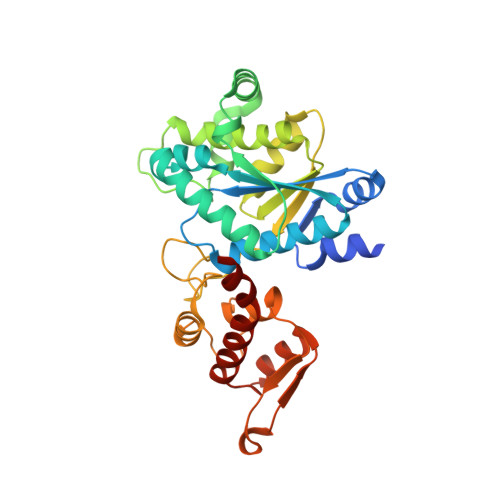
    5NSF

  • PubMed Abstract: 

    Allosteric information transfer in proteins has been linked to distinct vibrational energy transfer (VET) pathways in a number of theoretical studies. Experimental evidence for such pathways, however, is sparse because site-selective injection of vibrational energy into a protein, that is, localized heating, is required for their investigation. Here, we solved this problem by the site-specific incorporation of the non-canonical amino acid β-(1-azulenyl)-l-alanine (AzAla) through genetic code expansion. As an exception to Kasha's rule, AzAla undergoes ultrafast internal conversion and heating after S 1 excitation while upon S 2 excitation, it serves as a fluorescent label. We equipped PDZ3, a protein interaction domain of postsynaptic density protein 95, with this ultrafast heater at two distinct positions. We indeed observed VET from the incorporation sites in the protein to a bound peptide ligand on the picosecond timescale by ultrafast IR spectroscopy. This approach based on genetically encoded AzAla paves the way for detailed studies of VET and its role in a wide range of proteins.


  • Organizational Affiliation

    Institut für Chemie, Technische Universität Berlin, Müller-Breslau-Str. 10, 10623, Berlin, Germany.


Macromolecules
Find similar proteins by:  (by identity cutoff)  |  3D Structure
Entity ID: 1
MoleculeChains Sequence LengthOrganismDetailsImage
Tyrosine--tRNA ligaseA [auth B],
B [auth A],
C,
D
318Methanocaldococcus jannaschii DSM 2661Mutation(s): 0 
Gene Names: tyrSMJ0389
EC: 6.1.1.1
UniProt
Find proteins for Q57834 (Methanocaldococcus jannaschii (strain ATCC 43067 / DSM 2661 / JAL-1 / JCM 10045 / NBRC 100440))
Explore Q57834 
Go to UniProtKB:  Q57834
Entity Groups  
Sequence Clusters30% Identity50% Identity70% Identity90% Identity95% Identity100% Identity
UniProt GroupQ57834
Sequence Annotations
Expand
  • Reference Sequence
Small Molecules
Ligands 4 Unique
IDChains Name / Formula / InChI Key2D Diagram3D Interactions
96Z
Query on 96Z

Download Ideal Coordinates CCD File 
E [auth B],
H [auth A],
M [auth C],
N [auth D]
(2~{S})-2-azanyl-3-(2,6-dihydroazulen-1-yl)propanoic acid
C13 H15 N O2
HICXVJKDDWMICV-LBPRGKRZSA-N
PG4
Query on PG4

Download Ideal Coordinates CCD File 
L [auth A]TETRAETHYLENE GLYCOL
C8 H18 O5
UWHCKJMYHZGTIT-UHFFFAOYSA-N
GOL
Query on GOL

Download Ideal Coordinates CCD File 
F [auth B],
I [auth A]
GLYCEROL
C3 H8 O3
PEDCQBHIVMGVHV-UHFFFAOYSA-N
CA
Query on CA

Download Ideal Coordinates CCD File 
G [auth B],
J [auth A],
K [auth A]
CALCIUM ION
Ca
BHPQYMZQTOCNFJ-UHFFFAOYSA-N
Experimental Data & Validation

Experimental Data

  • Method: X-RAY DIFFRACTION
  • Resolution: 2.43 Å
  • R-Value Free: 0.240 
  • R-Value Work: 0.182 
  • R-Value Observed: 0.185 
  • Space Group: P 1
Unit Cell:
Length ( Å )Angle ( ˚ )
a = 56.14α = 85.7
b = 70.633β = 89.75
c = 77.624γ = 83.42
Software Package:
Software NamePurpose
PHENIXrefinement
XDSdata reduction
XDSdata scaling
PHASERphasing

Structure Validation

View Full Validation Report


Ligand Structure Quality Assessment 


Entry History 

Deposition Data

Revision History  (Full details and data files)

  • Version 1.0: 2019-01-16
    Type: Initial release
  • Version 1.1: 2019-02-13
    Changes: Data collection, Database references
  • Version 1.2: 2019-02-27
    Changes: Data collection, Database references
  • Version 1.3: 2019-10-16
    Changes: Data collection
  • Version 1.4: 2024-01-17
    Changes: Data collection, Database references, Derived calculations, Refinement description