5J37

Crystal structure of 60-mer BFDV Capsid Protein in complex with single stranded DNA


Experimental Data Snapshot

  • Method: X-RAY DIFFRACTION
  • Resolution: 2.30 Å
  • R-Value Free: 0.197 
  • R-Value Work: 0.173 
  • R-Value Observed: 0.175 

wwPDB Validation   3D Report Full Report


This is version 1.3 of the entry. See complete history


Literature

Structural insights into the assembly and regulation of distinct viral capsid complexes.

Sarker, S.Terron, M.C.Khandokar, Y.Aragao, D.Hardy, J.M.Radjainia, M.Jimenez-Zaragoza, M.de Pablo, P.J.Coulibaly, F.Luque, D.Raidal, S.R.Forwood, J.K.

(2016) Nat Commun 7: 13014-13014

  • DOI: https://doi.org/10.1038/ncomms13014
  • Primary Citation of Related Structures:  
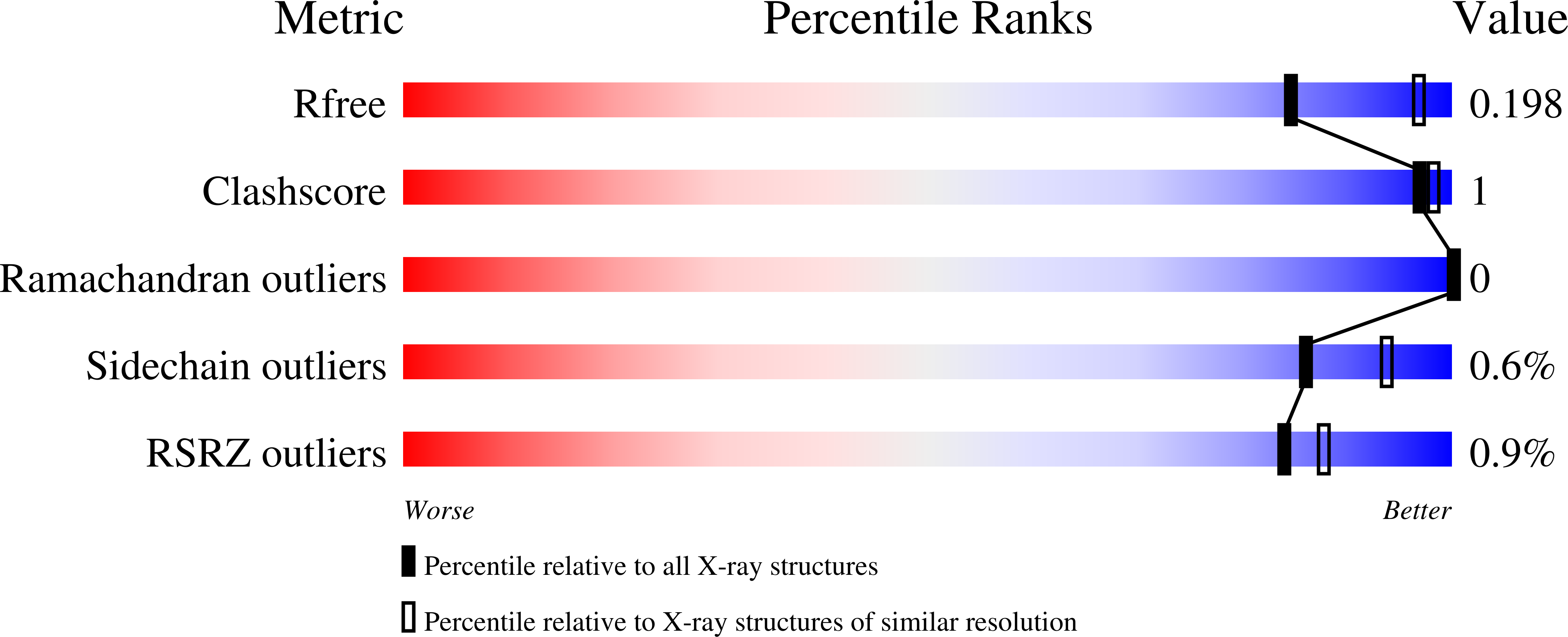
    5J09, 5J36, 5J37

  • PubMed Abstract: 

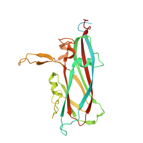
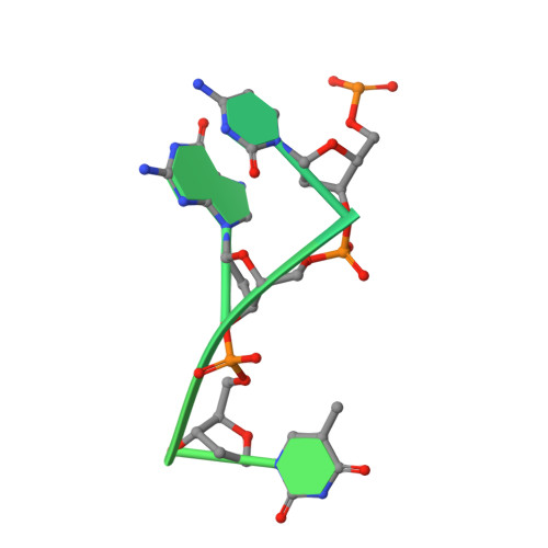
    The assembly and regulation of viral capsid proteins into highly ordered macromolecular complexes is essential for viral replication. Here, we utilize crystal structures of the capsid protein from the smallest and simplest known viruses capable of autonomously replicating in animal cells, circoviruses, to establish structural and mechanistic insights into capsid morphogenesis and regulation. The beak and feather disease virus, like many circoviruses, encode only two genes: a capsid protein and a replication initiation protein. The capsid protein forms distinct macromolecular assemblies during replication and here we elucidate these structures at high resolution, showing that these complexes reverse the exposure of the N-terminal arginine rich domain responsible for DNA binding and nuclear localization. We show that assembly of these complexes is regulated by single-stranded DNA (ssDNA), and provide a structural basis of capsid assembly around single-stranded DNA, highlighting novel binding interfaces distinct from the highly positively charged N-terminal ARM domain.


  • Organizational Affiliation

    School of Animal and Veterinary Sciences, Charles Sturt University, Boorooma Street, Wagga Wagga, New South Wales 2678, Australia.


Macromolecules

Find similar proteins by:  (by identity cutoff)  |  3D Structure
Entity ID: 1
MoleculeChains Sequence LengthOrganismDetailsImage
Beak and feather disease virus capsid proteinA [auth E],
C [auth A],
E [auth B],
G [auth C],
I [auth D]
257Beak and feather disease virusMutation(s): 0 
Gene Names: Cap
UniProt
Find proteins for A0A023R6W2 (Beak and feather disease virus)
Explore A0A023R6W2 
Go to UniProtKB:  A0A023R6W2
Entity Groups  
Sequence Clusters30% Identity50% Identity70% Identity90% Identity95% Identity100% Identity
UniProt GroupA0A023R6W2
Sequence Annotations
Expand
  • Reference Sequence
Find similar nucleic acids by:  (by identity cutoff)  |  3D Structure
Entity ID: 2
MoleculeChains LengthOrganismImage
single stranded DNAB [auth F],
D [auth G],
F [auth H],
H [auth I],
J
30synthetic construct
Sequence Annotations
Expand
  • Reference Sequence
Experimental Data & Validation

Experimental Data

  • Method: X-RAY DIFFRACTION
  • Resolution: 2.30 Å
  • R-Value Free: 0.197 
  • R-Value Work: 0.173 
  • R-Value Observed: 0.175 
  • Space Group: F 4 3 2
Unit Cell:
Length ( Å )Angle ( ˚ )
a = 377.28α = 90
b = 377.28β = 90
c = 377.28γ = 90
Software Package:
Software NamePurpose
PHENIXrefinement

Structure Validation

View Full Validation Report


Entry History 

Deposition Data

Revision History  (Full details and data files)

  • Version 1.0: 2016-05-04
    Type: Initial release
  • Version 1.1: 2016-10-12
    Changes: Database references
  • Version 1.2: 2023-03-29
    Changes: Data collection, Database references, Derived calculations, Structure summary
  • Version 1.3: 2023-10-25
    Changes: Data collection, Refinement description