5H4B

Crystal structure of Cbln4


Experimental Data Snapshot

  • Method: X-RAY DIFFRACTION
  • Resolution: 2.80 Å
  • R-Value Free: 0.233 
  • R-Value Work: 0.207 
  • R-Value Observed: 0.208 

wwPDB Validation   3D Report Full Report


Ligand Structure Quality Assessment 


This is version 1.3 of the entry. See complete history


Literature

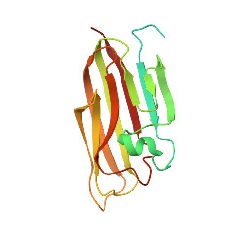
Cbln1 and Cbln4 Are Structurally Similar but Differ in GluD2 Binding Interactions.

Zhong, C.Shen, J.Zhang, H.Li, G.Shen, S.Wang, F.Hu, K.Cao, L.He, Y.Ding, J.

(2017) Cell Rep 20: 2328-2340

  • DOI: https://doi.org/10.1016/j.celrep.2017.08.031
  • Primary Citation of Related Structures:  
    5H48, 5H49, 5H4B, 5H4C

  • PubMed Abstract: 

    Unlike cerebellin 1 (Cbln1), which bridges neurexin (Nrxn) receptors and δ-type glutamate receptors in a trans-synaptic triad, Cbln4 was reported to have no or weak binding for the receptors despite sharing ∼70% sequence identity with Cbln1. Here, we report crystal structures of the homotrimers of the C1q domain of Cbln1 and Cbln4 at 2.2 and 2.3 Å resolution, respectively. Comparison of the structures suggests that the difference between Cbln1 and Cbln4 in GluD2 binding might be because of their sequence and structural divergence in loop CD. Surprisingly, we show that Cbln4 binds to Nrxn1β and forms a stable complex with the laminin, nectin, sex-hormone binding globulin (LNS) domain of Nrxn1β. Furthermore, the negative-stain electron microscopy reconstruction of hexameric full-length Cbln1 at 13 Å resolution and that of the Cbln4/Nrxn1β complex at 19 Å resolution suggest that Nrxn1β binds to the N-terminal region of Cbln4, probably through strand β10 of the S4 insert.


  • Organizational Affiliation

    National Center for Protein Science Shanghai, State Key Laboratory of Molecular Biology, Center for Excellence in Molecular Cell Science, Institute of Biochemistry and Cell Biology, University of Chinese Academy of Sciences, Chinese Academy of Sciences, 320 Yueyang Road, Shanghai 200031, P.R. China. Electronic address: czhong@sibcb.ac.cn.


Macromolecules
Find similar proteins by:  (by identity cutoff)  |  3D Structure
Entity ID: 1
MoleculeChains Sequence LengthOrganismDetailsImage
Cerebellin-4186Mus musculusMutation(s): 0 
Gene Names: Cbln4Cblnl1
UniProt
Find proteins for Q8BME9 (Mus musculus)
Explore Q8BME9 
Go to UniProtKB:  Q8BME9
Entity Groups  
Sequence Clusters30% Identity50% Identity70% Identity90% Identity95% Identity100% Identity
UniProt GroupQ8BME9
Sequence Annotations
Expand
  • Reference Sequence
Small Molecules
Ligands 1 Unique
IDChains Name / Formula / InChI Key2D Diagram3D Interactions
NAG
Query on NAG

Download Ideal Coordinates CCD File 
B [auth A]2-acetamido-2-deoxy-beta-D-glucopyranose
C8 H15 N O6
OVRNDRQMDRJTHS-FMDGEEDCSA-N
Experimental Data & Validation

Experimental Data

  • Method: X-RAY DIFFRACTION
  • Resolution: 2.80 Å
  • R-Value Free: 0.233 
  • R-Value Work: 0.207 
  • R-Value Observed: 0.208 
  • Space Group: P 43 3 2
Unit Cell:
Length ( Å )Angle ( ˚ )
a = 132.357α = 90
b = 132.357β = 90
c = 132.357γ = 90
Software Package:
Software NamePurpose
REFMACrefinement
HKL-3000data processing
HKL-3000data scaling
PHENIXphasing

Structure Validation

View Full Validation Report


Ligand Structure Quality Assessment 


Entry History & Funding Information

Deposition Data


Funding OrganizationLocationGrant Number
the National Natural Science Foundation of ChinaChina31370726
the Ministry of Science and Technology of ChinaChina2013CB910404

Revision History  (Full details and data files)

  • Version 1.0: 2017-09-13
    Type: Initial release
  • Version 1.1: 2017-09-20
    Changes: Database references
  • Version 1.2: 2018-01-17
    Changes: Database references
  • Version 1.3: 2020-07-29
    Type: Remediation
    Reason: Carbohydrate remediation
    Changes: Data collection, Derived calculations, Structure summary