5GH0

Crystal structure of the complex of bovine lactoperoxidase with mercaptoimidazole at 2.3 A resolution


Experimental Data Snapshot

  • Method: X-RAY DIFFRACTION
  • Resolution: 2.30 Å
  • R-Value Free: 0.194 
  • R-Value Work: 0.142 
  • R-Value Observed: 0.143 

wwPDB Validation   3D Report Full Report


Ligand Structure Quality Assessment 


This is version 2.1 of the entry. See complete history


Literature

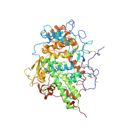
Design of anti-thyroid drugs: Binding studies and structure determination of the complex of lactoperoxidase with 2-mercaptoimidazole at 2.30 angstrom resolution

Sirohi, H.V.Singh, P.K.Iqbal, N.Sharma, P.Singh, A.K.Kaur, P.Sharma, S.Singh, T.P.

(2017) Proteins 85: 1882-1890

  • DOI: https://doi.org/10.1002/prot.25342
  • Primary Citation of Related Structures:  
    5GH0

  • PubMed Abstract: 

    Lactoperoxidase (LPO) belongs to mammalian heme peroxidase superfamily, which also includes myeloperoxidase (MPO), eosinophil peroxidase (EPO), and thyroid peroxidase (TPO). LPO catalyzes the oxidation of a number of substrates including thiocyanate while TPO catalyzes the biosynthesis of thyroid hormones. LPO is also been shown to catalyze the biosynthesis of thyroid hormones indicating similar functional and structural properties. The binding studies showed that 2-mercaptoimidazole (MZY) bound to LPO with a dissociation constant of 0.63 µM. The inhibition studies showed that the value of IC 50 was 17 µM. The crystal structure of the complex of LPO with MZY showed that MZY bound to LPO in the substrate-binding site on the distal heme side. MZY was oriented in the substrate-binding site in such a way that the sulfur atom is at a distance of 2.58 Å from the heme iron. Previously, a similar compound, 3-amino-1,2,4-triazole (amitrole) was also shown to bind to LPO in the substrate-binding site on the distal heme side. The amino nitrogen atom of amitrole occupied the same position as that of sulfur atom in the present structure indicating a similar mode of binding. Recently, the structure of the complex of LPO with a potent antithyroid drug, 1-methylimidazole-2-thiol (methimazole, MMZ) was also determined. It showed that MMZ bound to LPO in the substrate-binding site on the distal heme side with 2 orientations. The position of methyl group was same in the 2 orientations while the positions of sulfur atom differed indicating a higher preference for a methyl group.


  • Organizational Affiliation

    Department of Biophysics, All India Institute of Medical Sciences, New Delhi.


Macromolecules
Find similar proteins by:  (by identity cutoff)  |  3D Structure
Entity ID: 1
MoleculeChains Sequence LengthOrganismDetailsImage
Lactoperoxidase595Bos taurusMutation(s): 0 
EC: 1.11.1.7
UniProt
Find proteins for P80025 (Bos taurus)
Explore P80025 
Go to UniProtKB:  P80025
Entity Groups  
Sequence Clusters30% Identity50% Identity70% Identity90% Identity95% Identity100% Identity
UniProt GroupP80025
Sequence Annotations
Expand
  • Reference Sequence
Oligosaccharides

Help

Entity ID: 2
MoleculeChains Length2D Diagram Glycosylation3D Interactions
2-acetamido-2-deoxy-beta-D-glucopyranose-(1-4)-2-acetamido-2-deoxy-beta-D-glucopyranose
B
2N-Glycosylation
Glycosylation Resources
GlyTouCan:  G42666HT
GlyCosmos:  G42666HT
GlyGen:  G42666HT
Small Molecules
Ligands 6 Unique
IDChains Name / Formula / InChI Key2D Diagram3D Interactions
HEM
Query on HEM

Download Ideal Coordinates CCD File 
C [auth A]PROTOPORPHYRIN IX CONTAINING FE
C34 H32 Fe N4 O4
KABFMIBPWCXCRK-RGGAHWMASA-L
NAG
Query on NAG

Download Ideal Coordinates CCD File 
D [auth A],
E [auth A],
F [auth A]
2-acetamido-2-deoxy-beta-D-glucopyranose
C8 H15 N O6
OVRNDRQMDRJTHS-FMDGEEDCSA-N
MZY
Query on MZY

Download Ideal Coordinates CCD File 
K [auth A]1,3-dihydroimidazole-2-thione
C3 H4 N2 S
OXFSTTJBVAAALW-UHFFFAOYSA-N
NO3
Query on NO3

Download Ideal Coordinates CCD File 
G [auth A],
H [auth A]
NITRATE ION
N O3
NHNBFGGVMKEFGY-UHFFFAOYSA-N
CO3
Query on CO3

Download Ideal Coordinates CCD File 
I [auth A]CARBONATE ION
C O3
BVKZGUZCCUSVTD-UHFFFAOYSA-L
CA
Query on CA

Download Ideal Coordinates CCD File 
J [auth A]CALCIUM ION
Ca
BHPQYMZQTOCNFJ-UHFFFAOYSA-N
Modified Residues  1 Unique
IDChains TypeFormula2D DiagramParent
SEP
Query on SEP
A
L-PEPTIDE LINKINGC3 H8 N O6 PSER
Experimental Data & Validation

Experimental Data

  • Method: X-RAY DIFFRACTION
  • Resolution: 2.30 Å
  • R-Value Free: 0.194 
  • R-Value Work: 0.142 
  • R-Value Observed: 0.143 
  • Space Group: P 1 21 1
Unit Cell:
Length ( Å )Angle ( ˚ )
a = 54.63α = 90
b = 80.667β = 102.6
c = 77.68γ = 90
Software Package:
Software NamePurpose
REFMACrefinement
AUTOMARdata reduction
SCALEPACKdata scaling
MOLREPphasing

Structure Validation

View Full Validation Report


Ligand Structure Quality Assessment 


Entry History 

Deposition Data

Revision History  (Full details and data files)

  • Version 1.0: 2016-06-29
    Type: Initial release
  • Version 1.1: 2018-03-21
    Changes: Advisory, Database references, Derived calculations
  • Version 2.0: 2020-07-29
    Type: Remediation
    Reason: Carbohydrate remediation
    Changes: Atomic model, Data collection, Derived calculations, Structure summary
  • Version 2.1: 2023-11-08
    Changes: Data collection, Database references, Refinement description, Structure summary