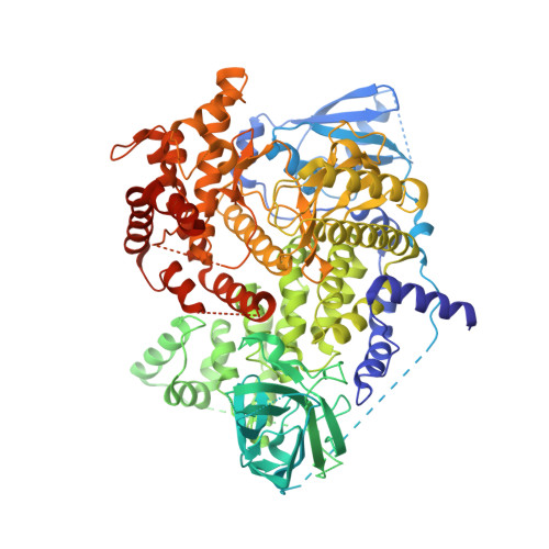
5G2N

X-ray structure of PI3Kinase Gamma in complex with Copanlisib


Experimental Data Snapshot

  • Method: X-RAY DIFFRACTION
  • Resolution: 2.68 Å
  • R-Value Free: 0.285 
  • R-Value Work: 0.211 
  • R-Value Observed: 0.215 

wwPDB Validation   3D Report Full Report


Ligand Structure Quality Assessment 


This is version 1.2 of the entry. See complete history


Literature

Discovery and Sar of Novel 2,3-Dihydroimidazo(1,2-C)Quinazoline Pi3K Inhibitors: Identification of Copanlisib (Bay 80-6946)

Schaefer, M.Scott, W.J.Hentemann, M.F.Rowley, R.B.Bull, C.O.Jenkins, S.Bullion, A.M.Johnson, J.Redman, A.Robbins, A.H.Esler, W.Fracasso, R.P.Garrison, T.Hamilton, M.Michels, M.Wood, J.E.Wilkie, D.P.Xiao, H.Levy, J.Liu, N.Stasik, E.Brands, M.Lefranc, J.

(2016) ChemMedChem 11: 1517

  • DOI: https://doi.org/10.1002/cmdc.201600148
  • Primary Citation of Related Structures:  
    5G2N

  • PubMed Abstract: 

    The phosphoinositide 3-kinase (PI3K) pathway is aberrantly activated in many disease states, including tumor cells, either by growth factor receptor tyrosine kinases or by the genetic mutation and amplification of key pathway components. A variety of PI3K isoforms play differential roles in cancers. As such, the development of PI3K inhibitors from novel compound classes should lead to differential pharmacological and pharmacokinetic profiles and allow exploration in various indications, combinations, and dosing regimens. A screening effort aimed at the identification of PI3Kγ inhibitors for the treatment of inflammatory diseases led to the discovery of the novel 2,3-dihydroimidazo[1,2-c]quinazoline class of PI3K inhibitors. A subsequent lead optimization program targeting cancer therapy focused on inhibition of PI3Kα and PI3Kβ. Herein, initial structure-activity relationship findings for this class and the optimization that led to the identification of copanlisib (BAY 80-6946) as a clinical candidate for the treatment of solid and hematological tumors are described.


  • Organizational Affiliation

    Global Development, Global Program Management, Bayer HealthCare Pharmaceuticals Inc., Whippany, NJ, 07981, USA. william.scott@bayer.com.


Macromolecules
Find similar proteins by:  (by identity cutoff)  |  3D Structure
Entity ID: 1
MoleculeChains Sequence LengthOrganismDetailsImage
PHOSPHATIDYLINOSITOL-4,5-BISPHOSPHATE 3-KINASE CATALYTIC SUBUNIT GAMMA ISOFORM980Homo sapiensMutation(s): 0 
EC: 2.7.1.137 (PDB Primary Data), 2.7.1.153 (PDB Primary Data)
UniProt & NIH Common Fund Data Resources
Find proteins for P48736 (Homo sapiens)
Explore P48736 
Go to UniProtKB:  P48736
PHAROS:  P48736
GTEx:  ENSG00000105851 
Entity Groups  
Sequence Clusters30% Identity50% Identity70% Identity90% Identity95% Identity100% Identity
UniProt GroupP48736
Sequence Annotations
Expand
  • Reference Sequence
Small Molecules
Ligands 2 Unique
IDChains Name / Formula / InChI Key2D Diagram3D Interactions
6E2
Query on 6E2

Download Ideal Coordinates CCD File 
B [auth A]2-azanyl-~{N}-[7-methoxy-8-(3-morpholin-4-ylpropoxy)-2,3-dihydroimidazo[1,2-c]quinazolin-5-yl]pyrimidine-5-carboxamide
C23 H28 N8 O4
PZBCKZWLPGJMAO-UHFFFAOYSA-N
SO4
Query on SO4

Download Ideal Coordinates CCD File 
C [auth A]SULFATE ION
O4 S
QAOWNCQODCNURD-UHFFFAOYSA-L
Binding Affinity Annotations 
IDSourceBinding Affinity
6E2 BindingDB:  5G2N IC50: min: 0.7, max: 6.4 (nM) from 3 assay(s)
Experimental Data & Validation

Experimental Data

  • Method: X-RAY DIFFRACTION
  • Resolution: 2.68 Å
  • R-Value Free: 0.285 
  • R-Value Work: 0.211 
  • R-Value Observed: 0.215 
  • Space Group: C 1 2 1
Unit Cell:
Length ( Å )Angle ( ˚ )
a = 144.762α = 90
b = 68.336β = 95.11
c = 106.472γ = 90
Software Package:
Software NamePurpose
REFMACrefinement
XDSdata reduction
XSCALEdata scaling

Structure Validation

View Full Validation Report


Ligand Structure Quality Assessment 


Entry History 

Revision History  (Full details and data files)

  • Version 1.0: 2016-04-20
    Type: Initial release
  • Version 1.1: 2016-06-29
    Changes: Database references
  • Version 1.2: 2016-08-10
    Changes: Database references