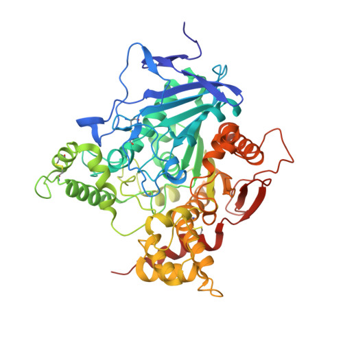
5FKJ

Crystal structure of mouse acetylcholinesterase in complex with C-547, an alkyl ammonium derivative of 6-methyl uracil


Experimental Data Snapshot

  • Method: X-RAY DIFFRACTION
  • Resolution: 3.13 Å
  • R-Value Free: 0.249 
  • R-Value Work: 0.207 
  • R-Value Observed: 0.208 

wwPDB Validation   3D Report Full Report


Ligand Structure Quality Assessment 


This is version 1.4 of the entry. See complete history


Literature

Slow-Binding Inhibition of Acetylcholinesterase by a 6-Methyluracil Alkyl-Ammonium Derivative: Mechanism and Advantages for Myasthenia Gravis Treatment.

Kharlamova, A.D.Lushchekina, S.V.Petrov, K.A.Kots, E.D.Nachon, F.V.Villard-Wandhammer, M.Zueva, I.V.Krejci, E.Reznik, V.S.Zobov, V.V.Nikolsky, E.E.Masson, P.

(2016) Biochem J 473: 1225

  • DOI: https://doi.org/10.1042/BCJ20160084
  • Primary Citation of Related Structures:  
    5FKJ

  • PubMed Abstract: 

    Inhibition of human AChE (acetylcholinesterase) and BChE (butyrylcholinesterase) by an alkylammonium derivative of 6-methyluracil, C-547, a potential drug for the treatment of MG (myasthenia gravis) was studied. Kinetic analysis of AChE inhibition showed that C-547 is a slow-binding inhibitor of type B, i.e. after formation of the initial enzyme·inhibitor complex (Ki=140 pM), an induced-fit step allows establishment of the final complex (Ki*=22 pM). The estimated koff is low, 0.05 min(-1) On the other hand, reversible inhibition of human BChE is a fast-binding process of mixed-type (Ki=1.77 μM; Ki'=3.17 μM). The crystal structure of mouse AChE complexed with C-547 was solved at 3.13 Å resolution. The complex is stabilized by cation-π, stacking and hydrogen-bonding interactions. Molecular dynamics simulations of the binding/dissociation processes of C-547 and C-35 (a non-charged analogue) to mouse and human AChEs were performed. Molecular modelling on mouse and human AChE showed that the slow step results from an enzyme conformational change that allows C-547 to cross the bottleneck in the active-site gorge, followed by formation of tight complex, as observed in the crystal structure. In contrast, the related non-charged compound C-35 is not a slow-binding inhibitor. It does not cross the bottleneck because it is not sensitive to the electrostatic driving force to reach the bottom of the gorge. Thus C-547 is one of the most potent and selective reversible inhibitors of AChE with a long residence time, τ=20 min, longer than for other reversible inhibitors used in the treatment of MG. This makes C-547 a promising drug for the treatment of this disease.


  • Organizational Affiliation

    A.E. Arbuzov Institute of Organic and Physical Chemistry of Russian Academy of Sciences, Arbuzov Str. 8, Kazan 420088 Russia.


Macromolecules
Find similar proteins by:  (by identity cutoff)  |  3D Structure
Entity ID: 1
MoleculeChains Sequence LengthOrganismDetailsImage
ACETYLCHOLINESTERASE
A, B, C, D
543Mus musculusMutation(s): 0 
EC: 3.1.1.7
UniProt & NIH Common Fund Data Resources
Find proteins for P21836 (Mus musculus)
Explore P21836 
Go to UniProtKB:  P21836
IMPC:  MGI:87876
Entity Groups  
Sequence Clusters30% Identity50% Identity70% Identity90% Identity95% Identity100% Identity
UniProt GroupP21836
Sequence Annotations
Expand
  • Reference Sequence
Small Molecules
Ligands 4 Unique
IDChains Name / Formula / InChI Key2D Diagram3D Interactions
G0W
Query on G0W

Download Ideal Coordinates CCD File 
E [auth A],
JA [auth D],
P [auth B],
X [auth C]
1,3-BIS[5(DIETHYL-O-NITROBENZYLAMMONIUM)PENTYL]-6-METHYLURACIL
C37 H56 N6 O6
WLTAFWKKNBLBHO-UHFFFAOYSA-N
NAG
Query on NAG

Download Ideal Coordinates CCD File 
F [auth A]
KA [auth D]
LA [auth D]
Q [auth B]
R [auth B]
F [auth A],
KA [auth D],
LA [auth D],
Q [auth B],
R [auth B],
Y [auth C]
2-acetamido-2-deoxy-beta-D-glucopyranose
C8 H15 N O6
OVRNDRQMDRJTHS-FMDGEEDCSA-N
SO4
Query on SO4

Download Ideal Coordinates CCD File 
IA [auth C],
SA [auth D],
W [auth B]
SULFATE ION
O4 S
QAOWNCQODCNURD-UHFFFAOYSA-L
CL
Query on CL

Download Ideal Coordinates CCD File 
AA [auth C]
BA [auth C]
CA [auth C]
DA [auth C]
EA [auth C]
AA [auth C],
BA [auth C],
CA [auth C],
DA [auth C],
EA [auth C],
FA [auth C],
G [auth A],
GA [auth C],
H [auth A],
HA [auth C],
I [auth A],
J [auth A],
K [auth A],
L [auth A],
M [auth A],
MA [auth D],
N [auth A],
NA [auth D],
O [auth A],
OA [auth D],
PA [auth D],
QA [auth D],
RA [auth D],
S [auth B],
T [auth B],
U [auth B],
V [auth B],
Z [auth C]
CHLORIDE ION
Cl
VEXZGXHMUGYJMC-UHFFFAOYSA-M
Experimental Data & Validation

Experimental Data

  • Method: X-RAY DIFFRACTION
  • Resolution: 3.13 Å
  • R-Value Free: 0.249 
  • R-Value Work: 0.207 
  • R-Value Observed: 0.208 
  • Space Group: P 21 21 21
Unit Cell:
Length ( Å )Angle ( ˚ )
a = 137.15α = 90
b = 175.45β = 90
c = 224.4γ = 90
Software Package:
Software NamePurpose
PHENIXrefinement
XDSdata reduction
XSCALEdata scaling
MOLREPphasing

Structure Validation

View Full Validation Report


Ligand Structure Quality Assessment 


Entry History 

Deposition Data

Revision History  (Full details and data files)

  • Version 1.0: 2016-03-16
    Type: Initial release
  • Version 1.1: 2016-05-11
    Changes: Database references
  • Version 1.2: 2017-08-09
    Changes: Data collection
  • Version 1.3: 2020-07-29
    Type: Remediation
    Reason: Carbohydrate remediation
    Changes: Data collection, Derived calculations, Other, Structure summary
  • Version 1.4: 2024-01-10
    Changes: Data collection, Database references, Refinement description, Structure summary