5F1C

Crystal structure of an invertebrate P2X receptor from the Gulf Coast tick in the presence of ATP and Zn2+ ion at 2.9 Angstroms


Experimental Data Snapshot

  • Method: X-RAY DIFFRACTION
  • Resolution: 2.90 Å
  • R-Value Free: 0.289 
  • R-Value Work: 0.255 
  • R-Value Observed: 0.256 

wwPDB Validation   3D Report Full Report


Ligand Structure Quality Assessment 


This is version 1.3 of the entry. See complete history


Literature

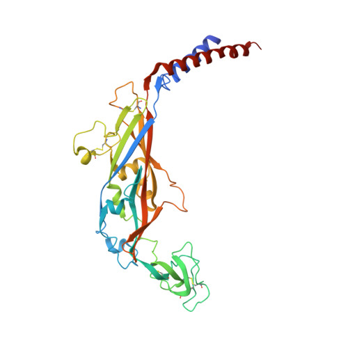
Structural Insights into Divalent Cation Modulations of ATP-Gated P2X Receptor Channels

Kasuya, G.Fujiwara, Y.Takemoto, M.Dohmae, N.Nakada-Nakura, Y.Ishitani, R.Hattori, M.Nureki, O.

(2016) Cell Rep 14: 932-944

  • DOI: https://doi.org/10.1016/j.celrep.2015.12.087
  • Primary Citation of Related Structures:  
    5F1C

  • PubMed Abstract: 

    P2X receptors are trimeric ATP-gated cation channels involved in physiological processes ranging widely from neurotransmission to pain and taste signal transduction. The modulation of the channel gating, including that by divalent cations, contributes to these diverse physiological functions of P2X receptors. Here, we report the crystal structure of an invertebrate P2X receptor from the Gulf Coast tick Amblyomma maculatum in the presence of ATP and Zn(2+) ion, together with electrophysiological and computational analyses. The structure revealed two distinct metal binding sites, M1 and M2, in the extracellular region. The M1 site, located at the trimer interface, is responsible for Zn(2+) potentiation by facilitating the structural change of the extracellular domain for pore opening. In contrast, the M2 site, coupled with the ATP binding site, might contribute to regulation by Mg(2+). Overall, our work provides structural insights into the divalent cation modulations of P2X receptors.


  • Organizational Affiliation

    Department of Biological Sciences, Graduate School of Science, The University of Tokyo, 2-11-16 Yayoi, Bunkyo-ku, Tokyo 113-0032, Japan.


Macromolecules
Find similar proteins by:  (by identity cutoff)  |  3D Structure
Entity ID: 1
MoleculeChains Sequence LengthOrganismDetailsImage
Putative uncharacterized protein
A, B, C
358Amblyomma maculatumMutation(s): 2 
Membrane Entity: Yes 
UniProt
Find proteins for G3MM57 (Amblyomma maculatum)
Explore G3MM57 
Go to UniProtKB:  G3MM57
Entity Groups  
Sequence Clusters30% Identity50% Identity70% Identity90% Identity95% Identity100% Identity
UniProt GroupG3MM57
Sequence Annotations
Expand
  • Reference Sequence
Small Molecules
Ligands 3 Unique
IDChains Name / Formula / InChI Key2D Diagram3D Interactions
ATP
Query on ATP

Download Ideal Coordinates CCD File 
L [auth A],
S [auth B],
T [auth C]
ADENOSINE-5'-TRIPHOSPHATE
C10 H16 N5 O13 P3
ZKHQWZAMYRWXGA-KQYNXXCUSA-N
NAG
Query on NAG

Download Ideal Coordinates CCD File 
D [auth A],
E [auth A],
M [auth B],
N [auth B],
U [auth C]
2-acetamido-2-deoxy-beta-D-glucopyranose
C8 H15 N O6
OVRNDRQMDRJTHS-FMDGEEDCSA-N
ZN
Query on ZN

Download Ideal Coordinates CCD File 
F [auth A]
G [auth A]
H [auth A]
I [auth A]
J [auth A]
F [auth A],
G [auth A],
H [auth A],
I [auth A],
J [auth A],
K [auth A],
O [auth B],
P [auth B],
Q [auth B],
R [auth B],
V [auth C],
W [auth C],
X [auth C]
ZINC ION
Zn
PTFCDOFLOPIGGS-UHFFFAOYSA-N
Experimental Data & Validation

Experimental Data

  • Method: X-RAY DIFFRACTION
  • Resolution: 2.90 Å
  • R-Value Free: 0.289 
  • R-Value Work: 0.255 
  • R-Value Observed: 0.256 
  • Space Group: P 21 21 21
Unit Cell:
Length ( Å )Angle ( ˚ )
a = 82.458α = 90
b = 139.9β = 90
c = 202.015γ = 90
Software Package:
Software NamePurpose
PHENIXrefinement
HKL-2000data scaling
PDB_EXTRACTdata extraction
HKL-2000data reduction
PHASERphasing

Structure Validation

View Full Validation Report


Ligand Structure Quality Assessment 


Entry History 

Deposition Data

Revision History  (Full details and data files)

  • Version 1.0: 2016-03-16
    Type: Initial release
  • Version 1.1: 2020-02-19
    Changes: Data collection, Derived calculations
  • Version 1.2: 2020-07-29
    Type: Remediation
    Reason: Carbohydrate remediation
    Changes: Data collection, Derived calculations, Structure summary
  • Version 1.3: 2023-11-08
    Changes: Data collection, Database references, Refinement description, Structure summary